6OIK | pdb_00006oik

Muscarinic acetylcholine receptor 2-Go complex


Experimental Data Snapshot

  • Method: ELECTRON MICROSCOPY
  • Resolution: 3.60 Å
  • Aggregation State: PARTICLE 
  • Reconstruction Method: SINGLE PARTICLE 

wwPDB Validation 3D Report Full Report

Validation slider image for 6OIK

This is version 1.4 of the entry. See complete history

Literature

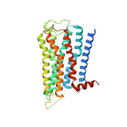
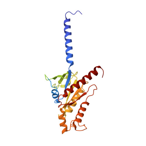
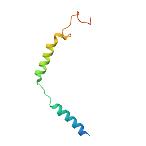
Structures of the M1 and M2 muscarinic acetylcholine receptor/G-protein complexes.

Maeda, S.Qu, Q.Robertson, M.J.Skiniotis, G.Kobilka, B.K.

(2019) Science 364: 552-557

  • DOI: https://doi.org/10.1126/science.aaw5188
  • Primary Citation Related Structures: 
    6OIJ, 6OIK

  • PubMed Abstract: 

    Muscarinic acetylcholine receptors are G protein-coupled receptors that respond to acetylcholine and play important signaling roles in the nervous system. There are five muscarinic receptor subtypes (M1R to M5R), which, despite sharing a high degree of sequence identity in the transmembrane region, couple to different heterotrimeric GTP-binding proteins (G proteins) to transmit signals. M1R, M3R, and M5R couple to the G q/ 11 family, whereas M2R and M4R couple to the G i/ o family. Here, we present and compare the cryo-electron microscopy structures of M1R in complex with G 11 and M2R in complex with G oA The M1R-G 11 complex exhibits distinct features, including an extended transmembrane helix 5 and carboxyl-terminal receptor tail that interacts with G protein. Detailed analysis of these structures provides a framework for understanding the molecular determinants of G-protein coupling selectivity.


  • Organizational Affiliation
    • Department of Molecular and Cellular Physiology, Stanford University School of Medicine, Stanford, CA, USA.

Macromolecule Content 

  • Total Structure Weight: 153.4 kDa 
  • Atom Count: 8,496 
  • Modeled Residue Count: 1,125 
  • Deposited Residue Count: 1,379 
  • Unique protein chains: 5

Macromolecules

Find similar proteins by:|  3D Structure
Entity ID: 1
MoleculeChains  Sequence LengthOrganismDetailsImage
Muscarinic acetylcholine receptor M2,Muscarinic acetylcholine receptor M2A [auth R]353Homo sapiensMutation(s): 3 
Gene Names: CHRM2
Membrane Entity: Yes 
UniProt & NIH Common Fund Data Resources
Find proteins for P08172 (Homo sapiens)
Explore P08172 
Go to UniProtKB:  P08172
PHAROS:  P08172
GTEx:  ENSG00000181072 
Entity Groups
Sequence Clusters30% Identity50% Identity70% Identity90% Identity95% Identity100% Identity
UniProt GroupP08172
Sequence Annotations
Expand
Reference Sequence
Find similar proteins by:|  3D Structure
Entity ID: 2
MoleculeChains  Sequence LengthOrganismDetailsImage
Guanine nucleotide-binding protein G(o) subunit alphaB [auth A]354Homo sapiensMutation(s): 4 
Gene Names: GNAO1
EC: 3.6.5
Membrane Entity: Yes 
UniProt & NIH Common Fund Data Resources
Find proteins for P09471 (Homo sapiens)
Explore P09471 
Go to UniProtKB:  P09471
PHAROS:  P09471
GTEx:  ENSG00000087258 
Entity Groups
Sequence Clusters30% Identity50% Identity70% Identity90% Identity95% Identity100% Identity
UniProt GroupP09471
Sequence Annotations
Expand
Reference Sequence
Find similar proteins by:|  3D Structure
Entity ID: 3
MoleculeChains  Sequence LengthOrganismDetailsImage
Guanine nucleotide-binding protein G(I)/G(S)/G(T) subunit beta-1C [auth B]345Homo sapiensMutation(s): 0 
Gene Names: GNB1
UniProt & NIH Common Fund Data Resources
Find proteins for P62873 (Homo sapiens)
Explore P62873 
Go to UniProtKB:  P62873
PHAROS:  P62873
GTEx:  ENSG00000078369 
Entity Groups
Sequence Clusters30% Identity50% Identity70% Identity90% Identity95% Identity100% Identity
UniProt GroupP62873
Sequence Annotations
Expand
Reference Sequence
Find similar proteins by:|  3D Structure
Entity ID: 4
MoleculeChains  Sequence LengthOrganismDetailsImage
Guanine nucleotide-binding protein G(I)/G(S)/G(O) subunit gamma-2D [auth G]71Homo sapiensMutation(s): 0 
Gene Names: GNG2
Membrane Entity: Yes 
UniProt & NIH Common Fund Data Resources
Find proteins for P59768 (Homo sapiens)
Explore P59768 
Go to UniProtKB:  P59768
PHAROS:  P59768
GTEx:  ENSG00000186469 
Entity Groups
Sequence Clusters30% Identity50% Identity70% Identity90% Identity95% Identity100% Identity
UniProt GroupP59768
Sequence Annotations
Expand
Reference Sequence
Find similar proteins by:|  3D Structure
Entity ID: 5
MoleculeChains  Sequence LengthOrganismDetailsImage
Antibody fragmentE [auth H]256Mus musculusMutation(s): 0 

Small Molecules

Ligands 2 Unique
IDChains Name / Formula / InChI Key2D Diagram3D Interactions
2CU

Query on 2CU


Download:Ideal Coordinates CCD File
G [auth R]3-amino-5-chloro-N-cyclopropyl-4-methyl-6-[2-(4-methylpiperazin-1-yl)-2-oxoethoxy]thieno[2,3-b]pyridine-2-carboxamide
C19 H24 Cl N5 O3 S
TYTGOXSAAQWLPJ-UHFFFAOYSA-N
IXO

Query on IXO


Download:Ideal Coordinates CCD File
F [auth R]4-(4,5-dihydro-1,2-oxazol-3-yloxy)-N,N,N-trimethylbut-2-yn-1-aminium
C10 H17 N2 O2
WXXOCGISBCTWPW-UHFFFAOYSA-N
Binding Affinity Annotations 
IDSourceBinding Affinity
2CU BindingDB:  6OIK Ki: min: 3548, max: 9333 (nM) from 5 assay(s)
IC50: 3.16e+4 (nM) from 1 assay(s)
IXO BindingDB:  6OIK Ki: min: 2.2, max: 257 (nM) from 6 assay(s)
EC50: min: 0.08, max: 0.28 (nM) from 3 assay(s)

Experimental Data & Validation

Experimental Data

  • Method: ELECTRON MICROSCOPY
  • Resolution: 3.60 Å
  • Aggregation State: PARTICLE 
  • Reconstruction Method: SINGLE PARTICLE 

Structure Validation

View Full Validation Report


Entry History 

& Funding Information

Deposition Data


Funding OrganizationLocationGrant Number
National Institutes of Health/National Institute of General Medical Sciences (NIH/NIGMS)United StatesR01GM083118

Revision History  (Full details and data files)

  • Version 1.0: 2019-05-08
    Type: Initial release
  • Version 1.1: 2019-05-22
    Changes: Data collection, Database references
  • Version 1.2: 2019-05-29
    Changes: Data collection, Database references
  • Version 1.3: 2020-01-08
    Changes: Author supporting evidence
  • Version 1.4: 2024-11-20
    Changes: Data collection, Database references, Structure summary