6OFC | pdb_00006ofc

Crystal structure of M. tuberculosis glutamine-dependent NAD+ synthetase complexed with Sulfonamide derivative 1, pyrophosphate, and glutamine


Experimental Data Snapshot

  • Method: X-RAY DIFFRACTION
  • Resolution: 3.14 Å
  • R-Value Free: 
    0.235 (Depositor), 0.217 (DCC) 
  • R-Value Work: 
    0.186 (Depositor) 
  • R-Value Observed: 
    0.189 (Depositor) 

Starting Model: experimental
View more details

wwPDB Validation 3D Report Full Report

Validation slider image for 6OFC

Ligand Structure Quality Assessment 


This is version 1.3 of the entry. See complete history

Literature

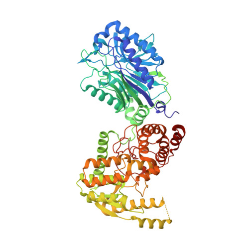
Different ways to transport ammonia in human and Mycobacterium tuberculosis NAD+synthetases.

Chuenchor, W.Doukov, T.I.Chang, K.T.Resto, M.Yun, C.S.Gerratana, B.

(2020) Nat Commun 11: 16-16

  • DOI: https://doi.org/10.1038/s41467-019-13845-4
  • Primary Citation Related Structures: 
    6OFB, 6OFC

  • PubMed Abstract: 

    NAD + synthetase is an essential enzyme of de novo and recycling pathways of NAD + biosynthesis in Mycobacterium tuberculosis but not in humans. This bifunctional enzyme couples the NAD + synthetase and glutaminase activities through an ammonia tunnel but free ammonia is also a substrate. Here we show that the Homo sapiens NAD + synthetase (hsNadE) lacks substrate specificity for glutamine over ammonia and displays a modest activation of the glutaminase domain compared to tbNadE. We report the crystal structures of hsNadE and NAD + synthetase from M. tuberculosis (tbNadE) with synthetase intermediate analogues. Based on the observed exclusive arrangements of the domains and of the intra- or inter-subunit tunnels we propose a model for the inter-domain communication mechanism for the regulation of glutamine-dependent activity and NH 3 transport. The structural and mechanistic comparison herein reported between hsNadE and tbNadE provides also a starting point for future efforts in the development of anti-TB drugs.


  • Organizational Affiliation
    • Departments of Chemistry and Biochemistry, University of Maryland, College Park, MD, 20742, USA.

Macromolecule Content 

  • Total Structure Weight: 303.5 kDa 
  • Atom Count: 20,893 
  • Modeled Residue Count: 2,655 
  • Deposited Residue Count: 2,716 
  • Unique protein chains: 1

Macromolecules

Find similar proteins by:|  3D Structure
Entity ID: 1
MoleculeChains  Sequence LengthOrganismDetailsImage
Glutamine-dependent NAD(+) synthetase
A, B, C, D
679Mycobacterium tuberculosis CDC1551Mutation(s): 1 
Gene Names: nadEMT2513
EC: 6.3.5.1
UniProt
Find proteins for P9WJJ3 (Mycobacterium tuberculosis (strain ATCC 25618 / H37Rv))
Explore P9WJJ3 
Go to UniProtKB:  P9WJJ3
Entity Groups
Sequence Clusters30% Identity50% Identity70% Identity90% Identity95% Identity100% Identity
UniProt GroupP9WJJ3
Sequence Annotations
Expand
Reference Sequence

Small Molecules

Ligands 5 Unique
IDChains Name / Formula / InChI Key2D Diagram3D Interactions
SFH
(Subject of Investigation/LOI)

Query on SFH


Download:Ideal Coordinates CCD File
E [auth A]
F [auth A]
G [auth A]
J [auth B]
K [auth B]
E [auth A],
F [auth A],
G [auth A],
J [auth B],
K [auth B],
L [auth B],
Q [auth C],
S [auth D]
5'-O-[(pyridine-3-carbonyl)sulfamoyl]adenosine
C16 H17 N7 O7 S
LBBNJCOHYSTAJW-UBEDBUPSSA-N
POP
(Subject of Investigation/LOI)

Query on POP


Download:Ideal Coordinates CCD File
N [auth B]PYROPHOSPHATE 2-
H2 O7 P2
XPPKVPWEQAFLFU-UHFFFAOYSA-L
GLN
(Subject of Investigation/LOI)

Query on GLN


Download:Ideal Coordinates CCD File
O [auth B],
R [auth C],
U [auth D]
GLUTAMINE
C5 H10 N2 O3
ZDXPYRJPNDTMRX-VKHMYHEASA-N
GOL

Query on GOL


Download:Ideal Coordinates CCD File
P [auth B],
V [auth D]
GLYCEROL
C3 H8 O3
PEDCQBHIVMGVHV-UHFFFAOYSA-N
CL

Query on CL


Download:Ideal Coordinates CCD File
H [auth A],
I [auth A],
M [auth B],
T [auth D]
CHLORIDE ION
Cl
VEXZGXHMUGYJMC-UHFFFAOYSA-M

Experimental Data & Validation

Experimental Data

  • Method: X-RAY DIFFRACTION
  • Resolution: 3.14 Å
  • R-Value Free:  0.235 (Depositor), 0.217 (DCC) 
  • R-Value Work:  0.186 (Depositor) 
  • R-Value Observed: 0.189 (Depositor) 
Space Group: P 41 21 2
Unit Cell:
Length ( Å )Angle ( ˚ )
a = 179.27α = 90
b = 179.27β = 90
c = 208.18γ = 90
Software Package:
Software NamePurpose
PHENIXrefinement
PHENIXrefinement
XDSdata reduction
SCALAdata scaling
PHENIXphasing

Structure Validation

View Full Validation Report


Ligand Structure Quality Assessment 


Entry History 

Deposition Data

Revision History  (Full details and data files)

  • Version 1.0: 2020-01-08
    Type: Initial release
  • Version 1.1: 2020-01-22
    Changes: Database references
  • Version 1.2: 2023-02-01
    Changes: Advisory, Database references
  • Version 1.3: 2023-10-25
    Changes: Data collection, Refinement description