6OAU | pdb_00006oau

Apo Structure of WT Lipoprotein Lipase in Complex with GPIHBP1 Mutant N78D N82D produced in GnTI-deficient HEK293-F cells


Experimental Data Snapshot

  • Method: X-RAY DIFFRACTION
  • Resolution: 2.48 Å
  • R-Value Free: 
    0.246 (Depositor), 0.250 (DCC) 
  • R-Value Work: 
    0.213 (Depositor), 0.218 (DCC) 
  • R-Value Observed: 
    0.215 (Depositor) 

Starting Model: experimental
View more details

wwPDB Validation 3D Report Full Report

Validation slider image for 6OAU

Ligand Structure Quality Assessment 


This is version 1.5 of the entry. See complete history

Literature

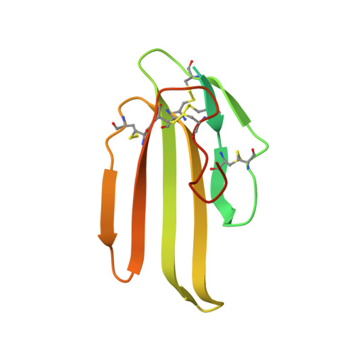
Structure of lipoprotein lipase in complex with GPIHBP1.

Arora, R.Nimonkar, A.V.Baird, D.Wang, C.Chiu, C.H.Horton, P.A.Hanrahan, S.Cubbon, R.Weldon, S.Tschantz, W.R.Mueller, S.Brunner, R.Lehr, P.Meier, P.Ottl, J.Voznesensky, A.Pandey, P.Smith, T.M.Stojanovic, A.Flyer, A.Benson, T.E.Romanowski, M.J.Trauger, J.W.

(2019) Proc Natl Acad Sci U S A 116: 10360-10365

  • DOI: https://doi.org/10.1073/pnas.1820171116
  • Primary Citation Related Structures: 
    6OAU, 6OAZ, 6OB0

  • PubMed Abstract: 

    Lipoprotein lipase (LPL) plays a central role in triglyceride (TG) metabolism. By catalyzing the hydrolysis of TGs present in TG-rich lipoproteins (TRLs), LPL facilitates TG utilization and regulates circulating TG and TRL concentrations. Until very recently, structural information for LPL was limited to homology models, presumably due to the propensity of LPL to unfold and aggregate. By coexpressing LPL with a soluble variant of its accessory protein glycosylphosphatidylinositol-anchored high-density lipoprotein binding protein 1 (GPIHBP1) and with its chaperone protein lipase maturation factor 1 (LMF1), we obtained a stable and homogenous LPL/GPIHBP1 complex that was suitable for structure determination. We report here X-ray crystal structures of human LPL in complex with human GPIHBP1 at 2.5-3.0 Å resolution, including a structure with a novel inhibitor bound to LPL. Binding of the inhibitor resulted in ordering of the LPL lid and lipid-binding regions and thus enabled determination of the first crystal structure of LPL that includes these important regions of the protein. It was assumed for many years that LPL was only active as a homodimer. The structures and additional biochemical data reported here are consistent with a new report that LPL, in complex with GPIHBP1, can be active as a monomeric 1:1 complex. The crystal structures illuminate the structural basis for LPL-mediated TRL lipolysis as well as LPL stabilization and transport by GPIHBP1.


  • Organizational Affiliation
    • Chemical Biology and Therapeutics, Novartis Institutes for Biomedical Research, Cambridge, MA 02139.

Macromolecule Content 

  • Total Structure Weight: 131.45 kDa 
  • Atom Count: 8,092 
  • Modeled Residue Count: 1,007 
  • Deposited Residue Count: 1,158 
  • Unique protein chains: 2

Macromolecules

Find similar proteins by:|  3D Structure
Entity ID: 1
MoleculeChains  Sequence LengthOrganismDetailsImage
Lipoprotein lipase
A, B
448Homo sapiensMutation(s): 0 
Gene Names: GPIHBP1HBP1
EC: 3.1.1.34 (PDB Primary Data), 3.1.1.32 (UniProt)
UniProt & NIH Common Fund Data Resources
Find proteins for P06858 (Homo sapiens)
Explore P06858 
Go to UniProtKB:  P06858
PHAROS:  P06858
GTEx:  ENSG00000175445 
Entity Groups
Sequence Clusters30% Identity50% Identity70% Identity90% Identity95% Identity100% Identity
UniProt GroupP06858
Glycosylation
Glycosylation Sites: 2Go to GlyGen: P06858-1
Sequence Annotations
Expand
Reference Sequence
Find similar proteins by:|  3D Structure
Entity ID: 2
MoleculeChains  Sequence LengthOrganismDetailsImage
Glycosylphosphatidylinositol-anchored high density lipoprotein-binding protein 1
C, D
131Homo sapiensMutation(s): 2 
Gene Names: GPIHBP1HBP1
UniProt & NIH Common Fund Data Resources
Find proteins for Q8IV16 (Homo sapiens)
Explore Q8IV16 
Go to UniProtKB:  Q8IV16
PHAROS:  Q8IV16
GTEx:  ENSG00000277494 
Entity Groups
Sequence Clusters30% Identity50% Identity70% Identity90% Identity95% Identity100% Identity
UniProt GroupQ8IV16
Sequence Annotations
Expand
Reference Sequence

Small Molecules

Ligands 3 Unique
IDChains Name / Formula / InChI Key2D Diagram3D Interactions
NAG

Query on NAG


Download:Ideal Coordinates CCD File
E [auth A],
F [auth A],
I [auth B],
J [auth B]
2-acetamido-2-deoxy-beta-D-glucopyranose
C8 H15 N O6
OVRNDRQMDRJTHS-FMDGEEDCSA-N
MLI

Query on MLI


Download:Ideal Coordinates CCD File
G [auth A]MALONATE ION
C3 H2 O4
OFOBLEOULBTSOW-UHFFFAOYSA-L
CA

Query on CA


Download:Ideal Coordinates CCD File
H [auth A],
K [auth B]
CALCIUM ION
Ca
BHPQYMZQTOCNFJ-UHFFFAOYSA-N

Experimental Data & Validation

Experimental Data

  • Method: X-RAY DIFFRACTION
  • Resolution: 2.48 Å
  • R-Value Free:  0.246 (Depositor), 0.250 (DCC) 
  • R-Value Work:  0.213 (Depositor), 0.218 (DCC) 
  • R-Value Observed: 0.215 (Depositor) 
Space Group: C 1 2 1
Unit Cell:
Length ( Å )Angle ( ˚ )
a = 177.48α = 90
b = 97.25β = 93.47
c = 77.21γ = 90
Software Package:
Software NamePurpose
BUSTERrefinement
Aimlessdata scaling
PDB_EXTRACTdata extraction
XDSdata reduction
BUSTERphasing

Structure Validation

View Full Validation Report


Ligand Structure Quality Assessment 


Entry History 

Deposition Data

Revision History  (Full details and data files)

  • Version 1.0: 2019-05-08
    Type: Initial release
  • Version 1.1: 2019-05-22
    Changes: Data collection, Database references
  • Version 1.2: 2019-06-05
    Changes: Data collection, Database references
  • Version 1.3: 2020-07-29
    Type: Remediation
    Reason: Carbohydrate remediation
    Changes: Data collection, Derived calculations, Structure summary
  • Version 1.4: 2023-10-11
    Changes: Advisory, Data collection, Database references, Refinement description, Structure summary
  • Version 1.5: 2024-11-20
    Changes: Structure summary