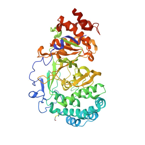
6OAL

Structure of human PARG complexed with JA2120


Experimental Data Snapshot

  • Method: X-RAY DIFFRACTION
  • Resolution: 1.60 Å
  • R-Value Free: 0.210 
  • R-Value Work: 0.181 
  • R-Value Observed: 0.182 

wwPDB Validation   3D Report Full Report


Ligand Structure Quality Assessment 


This is version 1.2 of the entry. See complete history


Literature

Selective small molecule PARG inhibitor causes replication fork stalling and cancer cell death.

Houl, J.H.Ye, Z.Brosey, C.A.Balapiti-Modarage, L.P.F.Namjoshi, S.Bacolla, A.Laverty, D.Walker, B.L.Pourfarjam, Y.Warden, L.S.Babu Chinnam, N.Moiani, D.Stegeman, R.A.Chen, M.K.Hung, M.C.Nagel, Z.D.Ellenberger, T.Kim, I.K.Jones, D.E.Ahmed, Z.Tainer, J.A.

(2019) Nat Commun 10: 5654-5654

  • DOI: https://doi.org/10.1038/s41467-019-13508-4
  • Primary Citation of Related Structures:  
    6O9X, 6O9Y, 6OA0, 6OA1, 6OA3, 6OAK, 6OAL

  • PubMed Abstract: 

    Poly(ADP-ribose)ylation (PARylation) by PAR polymerase 1 (PARP1) and PARylation removal by poly(ADP-ribose) glycohydrolase (PARG) critically regulate DNA damage responses; yet, conflicting reports obscure PARG biology and its impact on cancer cell resistance to PARP1 inhibitors. Here, we found that PARG expression is upregulated in many cancers. We employed chemical library screening to identify and optimize methylxanthine derivatives as selective bioavailable PARG inhibitors. Multiple crystal structures reveal how substituent positions on the methylxanthine core dictate binding modes and inducible-complementarity with a PARG-specific tyrosine clasp and arginine switch, supporting inhibitor specificity and a competitive inhibition mechanism. Cell-based assays show selective PARG inhibition and PARP1 hyperPARylation. Moreover, our PARG inhibitor sensitizes cells to radiation-induced DNA damage, suppresses replication fork progression and impedes cancer cell survival. In PARP inhibitor-resistant A172 glioblastoma cells, our PARG inhibitor shows comparable killing to Nedaplatin, providing further proof-of-concept that selectively inhibiting PARG can impair cancer cell survival.


  • Organizational Affiliation

    Departments of Cancer Biology and of Molecular and Cellular Oncology, University of Texas MD Anderson Cancer Center, 6767 Bertner Avenue, Houston, TX, 77030, USA.


Macromolecules
Find similar proteins by:  (by identity cutoff)  |  3D Structure
Entity ID: 1
MoleculeChains Sequence LengthOrganismDetailsImage
Poly(ADP-ribose) glycohydrolase529Homo sapiensMutation(s): 0 
Gene Names: PARG
EC: 3.2.1.143
UniProt & NIH Common Fund Data Resources
Find proteins for Q86W56 (Homo sapiens)
Explore Q86W56 
Go to UniProtKB:  Q86W56
PHAROS:  Q86W56
GTEx:  ENSG00000227345 
Entity Groups  
Sequence Clusters30% Identity50% Identity70% Identity90% Identity95% Identity100% Identity
UniProt GroupQ86W56
Sequence Annotations
Expand
  • Reference Sequence
Small Molecules
Ligands 1 Unique
IDChains Name / Formula / InChI Key2D Diagram3D Interactions
M0P
Query on M0P

Download Ideal Coordinates CCD File 
B [auth A]1,3-dimethyl-8-{[2-(morpholin-4-yl)ethyl]sulfanyl}-3,7-dihydro-1H-purine-2,6-dione
C13 H19 N5 O3 S
ZLIJABXMFRYUIA-UHFFFAOYSA-N
Modified Residues  1 Unique
IDChains TypeFormula2D DiagramParent
CME
Query on CME
A
L-PEPTIDE LINKINGC5 H11 N O3 S2CYS
Binding Affinity Annotations 
IDSourceBinding Affinity
M0P Binding MOAD:  6OAL IC50: 2.57e+4 (nM) from 1 assay(s)
Experimental Data & Validation

Experimental Data

  • Method: X-RAY DIFFRACTION
  • Resolution: 1.60 Å
  • R-Value Free: 0.210 
  • R-Value Work: 0.181 
  • R-Value Observed: 0.182 
  • Space Group: P 21 21 21
Unit Cell:
Length ( Å )Angle ( ˚ )
a = 66.302α = 90
b = 89.053β = 90
c = 94.29γ = 90
Software Package:
Software NamePurpose
PHENIXrefinement
XDSdata reduction
Aimlessdata scaling
PHASERphasing

Structure Validation

View Full Validation Report


Ligand Structure Quality Assessment 


Entry History & Funding Information

Deposition Data


Funding OrganizationLocationGrant Number
National Institutes of Health/National Cancer Institute (NIH/NCI)United States5R01CA200231-03

Revision History  (Full details and data files)

  • Version 1.0: 2020-03-18
    Type: Initial release
  • Version 1.1: 2020-05-27
    Changes: Database references
  • Version 1.2: 2023-10-11
    Changes: Data collection, Database references, Refinement description