6O9W | pdb_00006o9w

Crystal structure of Staphylococcus aureus BlaR1 antibiotic-sensor domain in complex with avibactam


Experimental Data Snapshot

  • Method: X-RAY DIFFRACTION
  • Resolution: 1.95 Å
  • R-Value Free: 
    0.225 (Depositor), 0.186 (DCC) 
  • R-Value Work: 
    0.185 (Depositor), 0.185 (DCC) 
  • R-Value Observed: 
    0.187 (Depositor) 

Starting Model: experimental
View more details

wwPDB Validation 3D Report Full Report

Validation slider image for 6O9W

Ligand Structure Quality Assessment 


This is version 1.3 of the entry. See complete history

Literature

Structural analysis of avibactam-mediated activation of the bla and mec divergons in methicillin-resistant Staphylococcus aureus .

Alexander, J.A.N.Radaeva, M.King, D.T.Chambers, H.F.Cherkasov, A.Chatterjee, S.S.Strynadka, N.C.J.

(2020) J Biological Chem 295: 10870-10884

  • DOI: https://doi.org/10.1074/jbc.RA120.013029
  • Primary Citation Related Structures: 
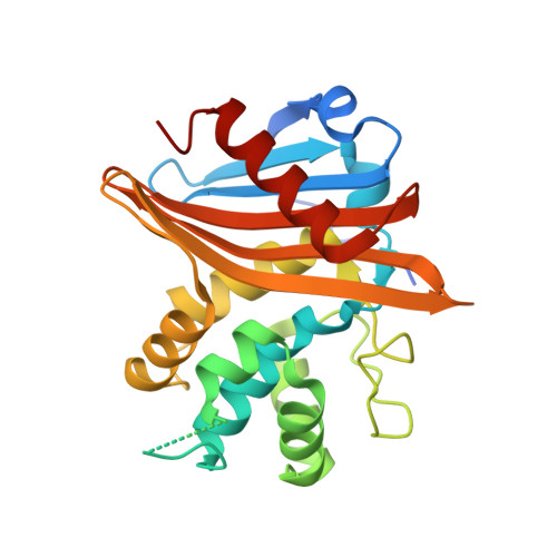
    6O9S, 6O9W

  • PubMed Abstract: 

    Methicillin-resistant Staphylococcus aureus (MRSA) infections cause significant mortality and morbidity globally. MRSA resistance to β-lactam antibiotics is mediated by two divergons that control levels of a β-lactamase, PC1, and a penicillin-binding protein poorly acylated by β-lactam antibiotics, PBP2a. Expression of genes encoding these proteins is controlled by two integral membrane proteins, BlaR1 and MecR1, which both have an extracellular β-lactam-binding sensor domain. Here, we solved the X-ray crystallographic structures of the BlaR1 and MecR1 sensor domains in complex with avibactam, a diazabicyclooctane β-lactamase inhibitor at 1.6-2.0 Å resolution. Additionally, we show that S. aureus SF8300, a clinically relevant strain from the USA300 clone of MRSA, responds to avibactam by up-regulating the expression of the blaZ and pbp2a antibiotic-resistance genes, encoding PC1 and PBP2a, respectively. The BlaR1-avibactam structure of the carbamoyl-enzyme intermediate revealed that avibactam is bound to the active-site serine in two orientations ∼180° to each other. Although a physiological role of the observed alternative pose remains to be validated, our structural results hint at the presence of a secondary sulfate-binding pocket that could be exploited in the design of future inhibitors of BlaR1/MecR1 sensor domains or the structurally similar class D β-lactamases. The MecR1-avibactam structure adopted a singular avibactam orientation similar to one of the two states observed in the BlaR1-avibactam structure. Given avibactam up-regulates expression of blaZ and pbp2a antibiotic resistance genes, we suggest further consideration and research is needed to explore what effects administering β-lactam-avibactam combinations have on treating MRSA infections.


  • Organizational Affiliation
    • Department of Biochemistry and Molecular Biology and Centre for Blood Research, The University of British Columbia, Vancouver, British Columbia, Canada.

Macromolecule Content 

  • Total Structure Weight: 61.37 kDa 
  • Atom Count: 4,151 
  • Modeled Residue Count: 472 
  • Deposited Residue Count: 518 
  • Unique protein chains: 1

Macromolecules

Find similar proteins by:|  3D Structure
Entity ID: 1
MoleculeChains  Sequence LengthOrganismDetailsImage
Regulatory protein BlaR1
A, B
259Staphylococcus aureusMutation(s): 0 
Gene Names: blaR1
UniProt
Find proteins for P18357 (Staphylococcus aureus)
Explore P18357 
Go to UniProtKB:  P18357
Entity Groups
Sequence Clusters30% Identity50% Identity70% Identity90% Identity95% Identity100% Identity
UniProt GroupP18357
Sequence Annotations
Expand
Reference Sequence

Small Molecules

Ligands 1 Unique
IDChains Name / Formula / InChI Key2D Diagram3D Interactions
NXL

Query on NXL


Download:Ideal Coordinates CCD File
C [auth A],
D [auth B]
(2S,5R)-1-formyl-5-[(sulfooxy)amino]piperidine-2-carboxamide
C7 H13 N3 O6 S
WJDGWXPPFHLLNL-RITPCOANSA-N

Experimental Data & Validation

Experimental Data

  • Method: X-RAY DIFFRACTION
  • Resolution: 1.95 Å
  • R-Value Free:  0.225 (Depositor), 0.186 (DCC) 
  • R-Value Work:  0.185 (Depositor), 0.185 (DCC) 
  • R-Value Observed: 0.187 (Depositor) 
Space Group: P 1 21 1
Unit Cell:
Length ( Å )Angle ( ˚ )
a = 53.355α = 90
b = 92.647β = 104.57
c = 56.472γ = 90
Software Package:
Software NamePurpose
PHENIXrefinement
XDSdata reduction
Aimlessdata scaling
PHASERphasing
PDB_EXTRACTdata extraction

Structure Validation

View Full Validation Report


Ligand Structure Quality Assessment 


Entry History 

& Funding Information

Deposition Data


Funding OrganizationLocationGrant Number
Canadian Institutes of Health Research (CIHR)Canada--

Revision History  (Full details and data files)

  • Version 1.0: 2020-06-10
    Type: Initial release
  • Version 1.1: 2020-10-07
    Changes: Database references
  • Version 1.2: 2023-10-11
    Changes: Data collection, Database references, Refinement description
  • Version 1.3: 2024-10-16
    Changes: Structure summary