6O9U | pdb_00006o9u

KirBac3.1 at a resolution of 2 Angstroms


Experimental Data Snapshot

  • Method: X-RAY DIFFRACTION
  • Resolution: 2.00 Å
  • R-Value Free: 
    0.227 (Depositor), 0.229 (DCC) 
  • R-Value Work: 
    0.202 (Depositor) 
  • R-Value Observed: 
    0.203 (Depositor) 

Starting Model: experimental
View more details

wwPDB Validation 3D Report Full Report

Validation slider image for 6O9U

Ligand Structure Quality Assessment 


This is version 1.3 of the entry. See complete history. 

Literature

A constricted opening in Kir channels does not impede potassium conduction.

Black, K.A., He, S., Jin, R., Miller, D.M., Bolla, J.R., Clarke, O.B., Johnson, P., Windley, M., Burns, C.J., Hill, A.P., Laver, D., Robinson, C.V., Smith, B.J., Gulbis, J.M.

(2020) Nat Commun 11: 3024-3024

  • DOI: https://doi.org/10.1038/s41467-020-16842-0
  • Primary Citation Related Structures: 
    6O9T, 6O9U, 6O9V

  • PubMed Abstract: 

    The canonical mechanistic model explaining potassium channel gating is of a conformational change that alternately dilates and constricts a collar-like intracellular entrance to the pore. It is based on the premise that K + ions maintain a complete hydration shell while passing between the transmembrane cavity and cytosol, which must be accommodated. To put the canonical model to the test, we locked the conformation of a Kir K + channel to prevent widening of the narrow collar. Unexpectedly, conduction was unimpaired in the locked channels. In parallel, we employed all-atom molecular dynamics to simulate K + ions moving along the conduction pathway between the lower cavity and cytosol. During simulations, the constriction did not significantly widen. Instead, transient loss of some water molecules facilitated K + permeation through the collar. The low free energy barrier to partial dehydration in the absence of conformational change indicates Kir channels are not gated by the canonical mechanism.


  • Organizational Affiliation: 
    • Structural Biology Division, The Walter and Eliza Hall Institute of Medical Research, Parkville, VIC, 3052, Australia.

Macromolecule Content 

  • Total Structure Weight: 36.37 kDa 
  • Atom Count: 2,473 
  • Modeled Residue Count: 283 
  • Deposited Residue Count: 301 
  • Unique protein chains: 1

Macromolecules

Find similar proteins by:|  3D Structure
Entity ID: 1
MoleculeChains  Sequence LengthOrganismDetailsImage
Inward rectifier potassium channel Kirbac3.1301Paramagnetospirillum magnetotacticumMutation(s): 0 
Membrane Entity: Yes 
UniProt
Find proteins for D9N164 (Paramagnetospirillum magnetotacticum)
Explore D9N164 
Go to UniProtKB:  D9N164
Entity Groups
Sequence Clusters30% Identity50% Identity70% Identity90% Identity95% Identity100% Identity
UniProt GroupD9N164
Sequence Annotations
Expand
Reference Sequence

Small Molecules

Ligands 6 Unique
IDChains Name / Formula / InChI Key2D Diagram3D Interactions
PCW

Query on PCW


Download:Ideal Coordinates CCD File
P [auth A],
Q [auth A]
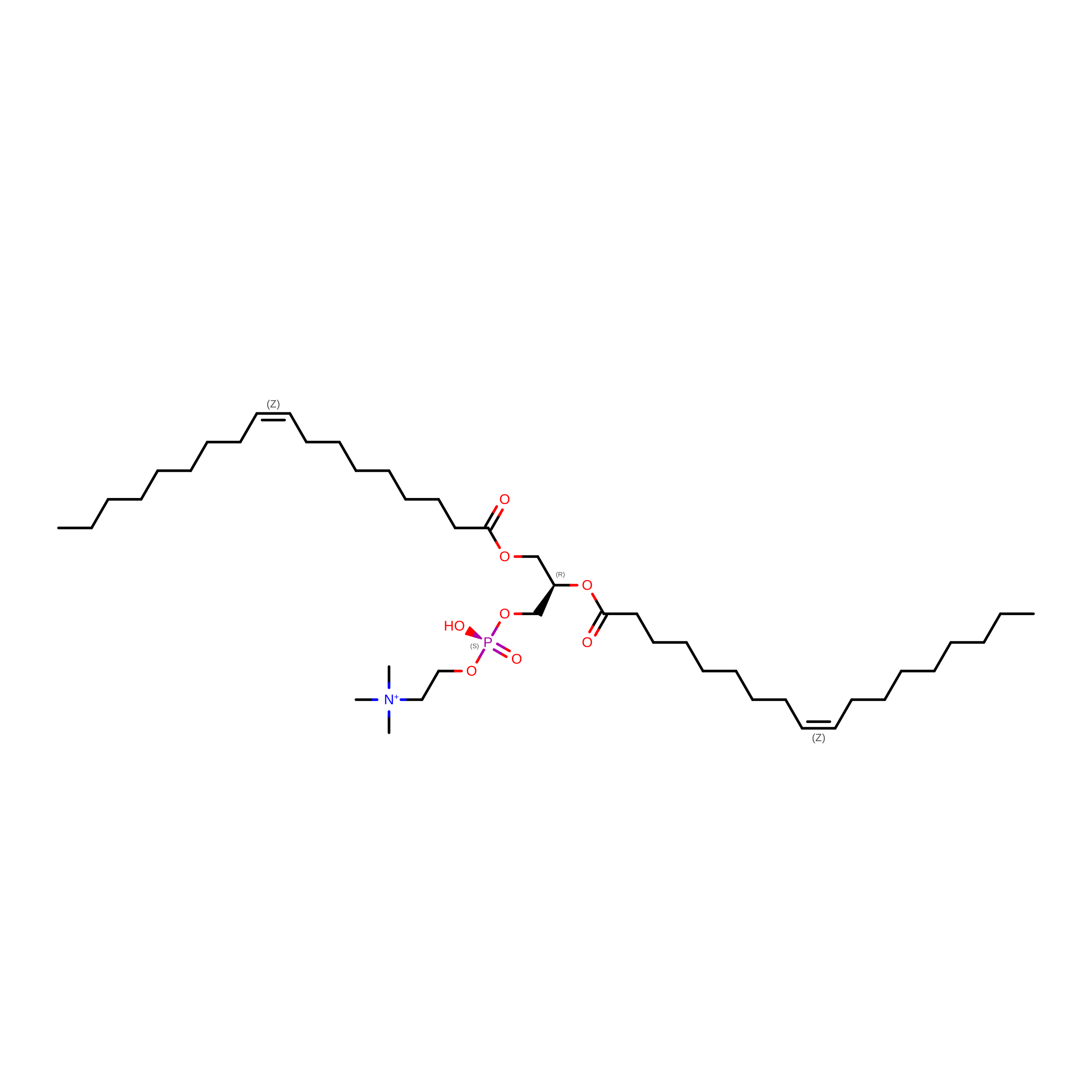
1,2-DIOLEOYL-SN-GLYCERO-3-PHOSPHOCHOLINE
C44 H85 N O8 P
SNKAWJBJQDLSFF-NVKMUCNASA-O
Z3P

Query on Z3P


Download:Ideal Coordinates CCD File
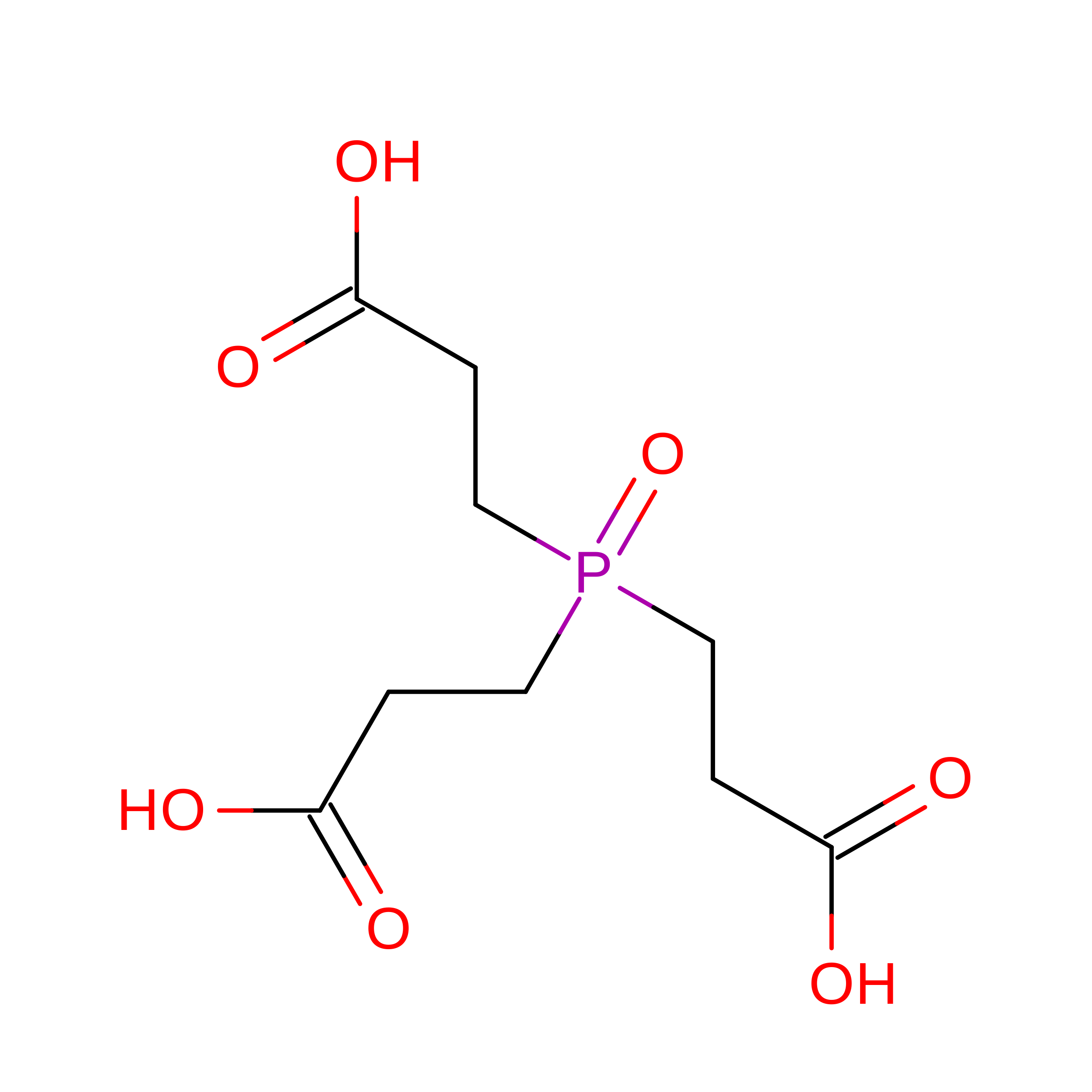
R [auth A]3,3',3''-phosphoryltripropanoic acid
C9 H15 O7 P
XJGZCDJZBUBTKW-UHFFFAOYSA-N
BA

Query on BA


Download:Ideal Coordinates CCD File
N [auth A]BARIUM ION
Ba
XDFCIPNJCBUZJN-UHFFFAOYSA-N
TMO

Query on TMO


Download:Ideal Coordinates CCD File
B [auth A],
C [auth A],
D [auth A]
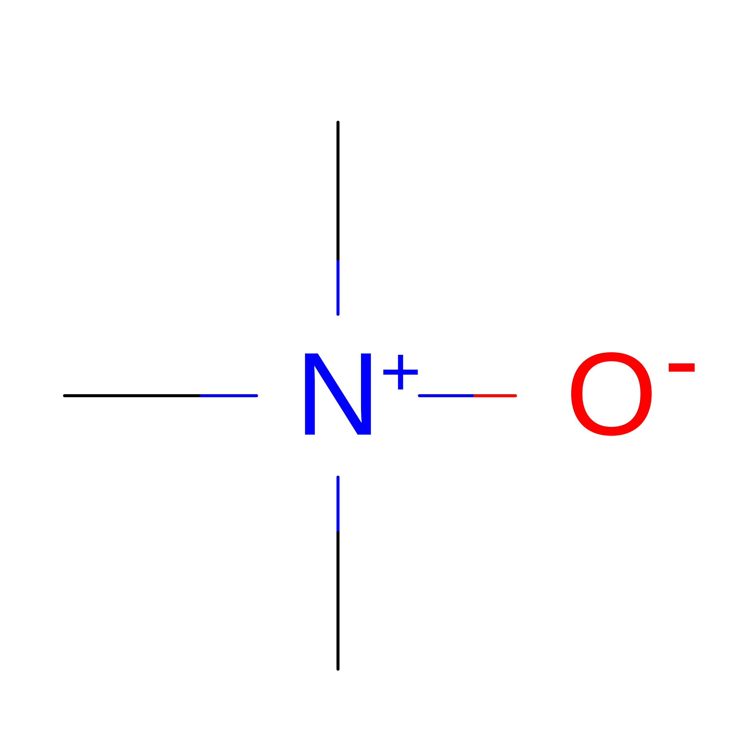
trimethylamine oxide
C3 H9 N O
UYPYRKYUKCHHIB-UHFFFAOYSA-N
K

Query on K


Download:Ideal Coordinates CCD File
E [auth A]
F [auth A]
G [auth A]
H [auth A]
I [auth A]
E [auth A],
F [auth A],
G [auth A],
H [auth A],
I [auth A],
J [auth A],
K [auth A],
L [auth A],
M [auth A]
POTASSIUM ION
K
NPYPAHLBTDXSSS-UHFFFAOYSA-N
CL

Query on CL


Download:Ideal Coordinates CCD File
O [auth A]CHLORIDE ION
Cl
VEXZGXHMUGYJMC-UHFFFAOYSA-M

Experimental Data & Validation

Experimental Data

  • Method: X-RAY DIFFRACTION
  • Resolution: 2.00 Å
  • R-Value Free:  0.227 (Depositor), 0.229 (DCC) 
  • R-Value Work:  0.202 (Depositor) 
  • R-Value Observed: 0.203 (Depositor) 
Space Group: P 4 21 2
Unit Cell:
Length ( Å )Angle ( ˚ )
a = 105.955α = 90
b = 105.955β = 90
c = 89.639γ = 90
Software Package:
Software NamePurpose
PHENIXrefinement
PDB_EXTRACTdata extraction
DENZOdata reduction
SCALEPACKdata scaling
PHASERphasing

Structure Validation

View Full Validation Report


Ligand Structure Quality Assessment 


Entry History 

& Funding Information

Deposition Data


Funding OrganizationLocationGrant Number
National Health and Medical Research Council (NHMRC, Australia)Australia--

Revision History  (Full details and data files)

  • Version 1.0: 2020-05-27
    Type: Initial release
  • Version 1.1: 2020-12-09
    Changes: Database references, Derived calculations
  • Version 1.2: 2020-12-16
    Changes: Database references
  • Version 1.3: 2023-10-11
    Changes: Data collection, Database references, Refinement description