6O56 | pdb_00006o56

HNH Nuclease from S. pyogenes Cas9


Experimental Data Snapshot

  • Method: X-RAY DIFFRACTION
  • Resolution: 1.90 Å
  • R-Value Free: 
    0.253 (Depositor), 0.251 (DCC) 
  • R-Value Work: 
    0.205 (Depositor), 0.205 (DCC) 
  • R-Value Observed: 
    0.207 (Depositor) 

Starting Model: experimental
View more details

wwPDB Validation 3D Report Full Report

Validation slider image for 6O56

This is version 1.3 of the entry. See complete history

Literature

Allosteric Motions of the CRISPR-Cas9 HNH Nuclease Probed by NMR and Molecular Dynamics.

East, K.W.Newton, J.C.Morzan, U.N.Narkhede, Y.B.Acharya, A.Skeens, E.Jogl, G.Batista, V.S.Palermo, G.Lisi, G.P.

(2020) J Am Chem Soc 142: 1348-1358

  • DOI: https://doi.org/10.1021/jacs.9b10521
  • Primary Citation Related Structures: 
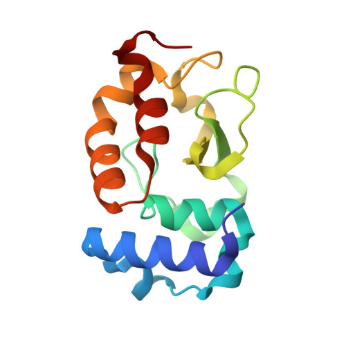
    6O56

  • PubMed Abstract: 

    CRISPR-Cas9 is a widely employed genome-editing tool with functionality reliant on the ability of the Cas9 endonuclease to introduce site-specific breaks in double-stranded DNA. In this system, an intriguing allosteric communication has been suggested to control its DNA cleavage activity through flexibility of the catalytic HNH domain. Here, solution NMR experiments and a novel Gaussian-accelerated molecular dynamics (GaMD) simulation method are used to capture the structural and dynamic determinants of allosteric signaling within the HNH domain. We reveal the existence of a millisecond time scale dynamic pathway that spans HNH from the region interfacing the adjacent RuvC nuclease and propagates up to the DNA recognition lobe in full-length CRISPR-Cas9. These findings reveal a potential route of signal transduction within the CRISPR-Cas9 HNH nuclease, advancing our understanding of the allosteric pathway of activation. Further, considering the role of allosteric signaling in the specificity of CRISPR-Cas9, this work poses the mechanistic basis for novel engineering efforts aimed at improving its genome-editing capability.


  • Organizational Affiliation
    • Department of Molecular Biology, Cell Biology & Biochemistry , Brown University , Providence , Rhode Island 02903 , United States.

Macromolecule Content 

  • Total Structure Weight: 31.97 kDa 
  • Atom Count: 2,525 
  • Modeled Residue Count: 270 
  • Deposited Residue Count: 270 
  • Unique protein chains: 1

Macromolecules

Find similar proteins by:|  3D Structure
Entity ID: 1
MoleculeChains  Sequence LengthOrganismDetailsImage
CRISPR-associated endonuclease Cas9/Csn1
A, B
135Streptococcus pyogenes serotype M1Mutation(s): 0 
Gene Names: cas9csn1SPy_1046
EC: 3.1
UniProt
Find proteins for Q99ZW2 (Streptococcus pyogenes serotype M1)
Explore Q99ZW2 
Go to UniProtKB:  Q99ZW2
Entity Groups
Sequence Clusters30% Identity50% Identity70% Identity90% Identity95% Identity100% Identity
UniProt GroupQ99ZW2
Sequence Annotations
Expand
Reference Sequence

Experimental Data & Validation

Experimental Data

  • Method: X-RAY DIFFRACTION
  • Resolution: 1.90 Å
  • R-Value Free:  0.253 (Depositor), 0.251 (DCC) 
  • R-Value Work:  0.205 (Depositor), 0.205 (DCC) 
  • R-Value Observed: 0.207 (Depositor) 
Space Group: P 21 21 21
Unit Cell:
Length ( Å )Angle ( ˚ )
a = 51.525α = 90
b = 56.063β = 90
c = 80.669γ = 90
Software Package:
Software NamePurpose
PHENIXrefinement
XDSdata reduction
Aimlessdata scaling
PHASERphasing
PDB_EXTRACTdata extraction

Structure Validation

View Full Validation Report


Entry History 

& Funding Information

Deposition Data


Funding OrganizationLocationGrant Number
Other privateUnited States--

Revision History  (Full details and data files)

  • Version 1.0: 2020-01-15
    Type: Initial release
  • Version 1.1: 2020-01-22
    Changes: Database references
  • Version 1.2: 2020-02-05
    Changes: Database references
  • Version 1.3: 2023-10-11
    Changes: Data collection, Database references, Refinement description