6O4M | pdb_00006o4m

Racemic melittin


Experimental Data Snapshot

  • Method: X-RAY DIFFRACTION
  • Resolution: 1.27 Å
  • R-Value Free: 
    0.235 (Depositor), 0.235 (DCC) 
  • R-Value Work: 
    0.201 (Depositor), 0.200 (DCC) 

Starting Model: experimental
View more details

wwPDB Validation 3D Report Full Report

Validation slider image for 6O4M

This is version 1.5 of the entry. See complete history

Literature

Retention of Native Quaternary Structure in Racemic Melittin Crystals.

Kurgan, K.W.Kleman, A.F.Bingman, C.A.Kreitler, D.F.Weisblum, B.Forest, K.T.Gellman, S.H.

(2019) J Am Chem Soc 141: 7704-7708

  • DOI: https://doi.org/10.1021/jacs.9b02691
  • Primary Citation Related Structures: 
    6O4M

  • PubMed Abstract: 

    Racemic crystallography has been used to elucidate the secondary and tertiary structures of peptides and small proteins that are recalcitrant to conventional crystallization. It is unclear, however, whether racemic crystallography can capture native quaternary structure, which could be disrupted by heterochiral associations. We are exploring the use of racemic crystallography to characterize the self-assembly behavior of membrane-associated peptides, very few of which have been crystallized. We report a racemic crystal structure of the membrane-active peptide melittin; the new structure allows comparison with a previously reported crystal structure of L-melittin. The tetrameric assembly observed in crystalline L-melittin has been proposed to represent the tetrameric state detected in solution for this peptide. This tetrameric assembly is precisely reproduced in the racemic crystal, which strengthens the conclusion that the tetramer is biologically relevant. More broadly, these findings suggest that racemic crystallography can provide insight on native quaternary structure.


  • Organizational Affiliation
    • Department of Structural Biology, Jacobs School of Medicine and Biomedical Sciences , University at Buffalo , Buffalo , New York 14203-1102 , United States.

Macromolecule Content 

  • Total Structure Weight: 12.26 kDa 
  • Atom Count: 1,040 
  • Modeled Residue Count: 108 
  • Deposited Residue Count: 108 
  • Unique protein chains: 2

Macromolecules

Find similar proteins by:|  3D Structure
Entity ID: 1
MoleculeChains  Sequence LengthOrganismDetailsImage
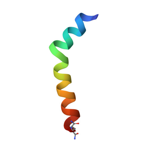
D-MelittinA [auth D],
D [auth C]
27Apis melliferaMutation(s): 1 
UniProt
Find proteins for P01501 (Apis mellifera)
Explore P01501 
Go to UniProtKB:  P01501
Entity Groups
UniProt GroupP01501
Sequence Annotations
Expand
Reference Sequence
Find similar proteins by:|  3D Structure
Entity ID: 2
MoleculeChains  Sequence LengthOrganismDetailsImage
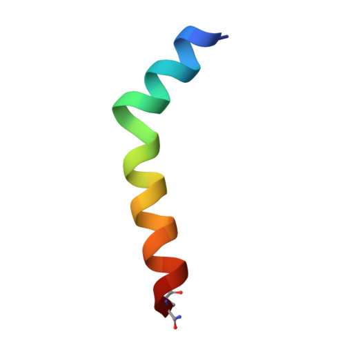
MelittinB [auth A],
C [auth B]
27Apis melliferaMutation(s): 1 
UniProt
Find proteins for P01501 (Apis mellifera)
Explore P01501 
Go to UniProtKB:  P01501
Entity Groups
Sequence Clusters30% Identity50% Identity70% Identity90% Identity95% Identity100% Identity
UniProt GroupP01501
Sequence Annotations
Expand
Reference Sequence

Small Molecules

Ligands 1 Unique
IDChains Name / Formula / InChI Key2D Diagram3D Interactions
SO4

Query on SO4


Download:Ideal Coordinates CCD File
E [auth D]
F [auth D]
G [auth A]
H [auth A]
I [auth B]
E [auth D],
F [auth D],
G [auth A],
H [auth A],
I [auth B],
J [auth B],
K [auth B],
L [auth C],
M [auth C]
SULFATE ION
O4 S
QAOWNCQODCNURD-UHFFFAOYSA-L
Modified Residues  11 Unique
IDChains TypeFormula2D DiagramParent
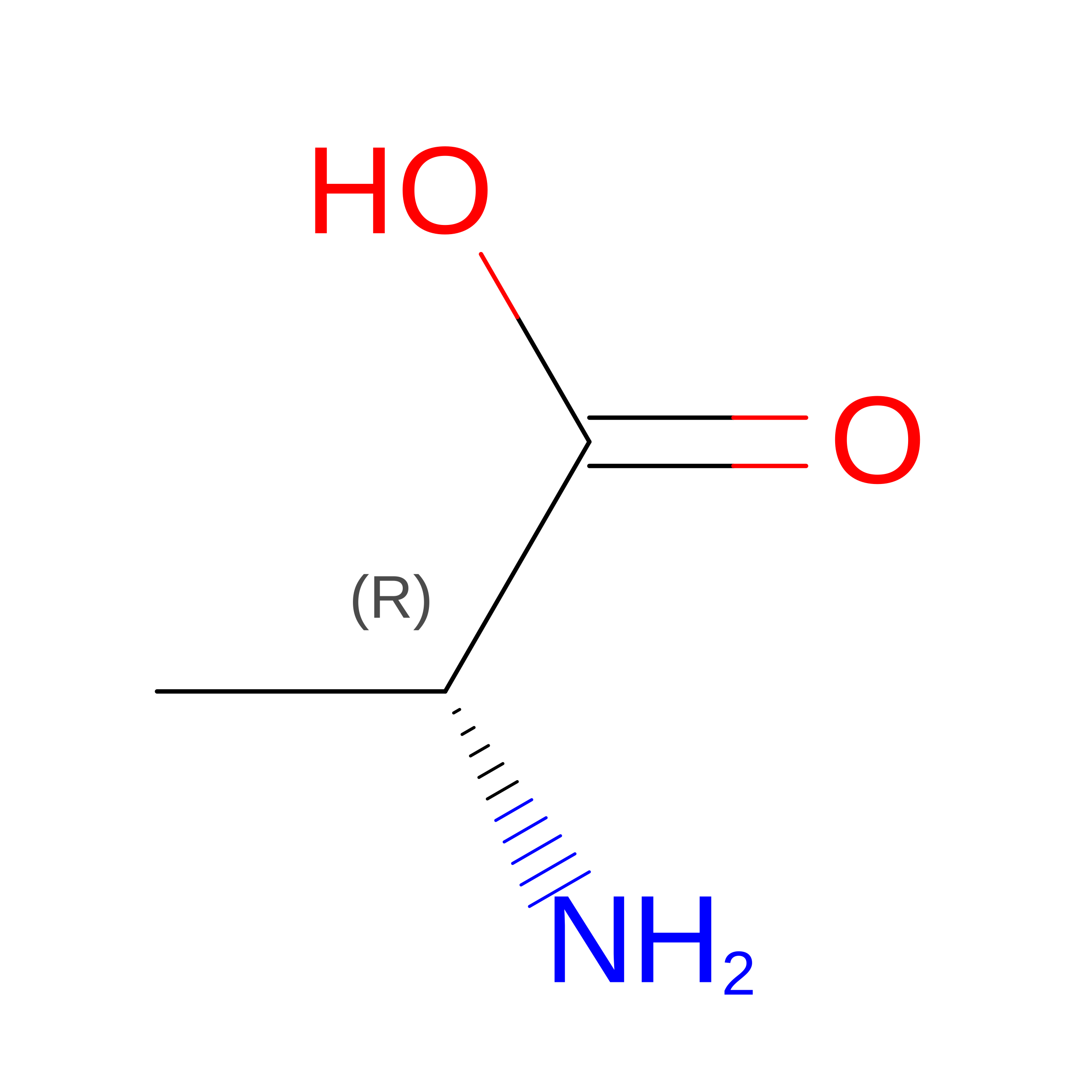
DAL
Query on DAL
A [auth D],
D [auth C]
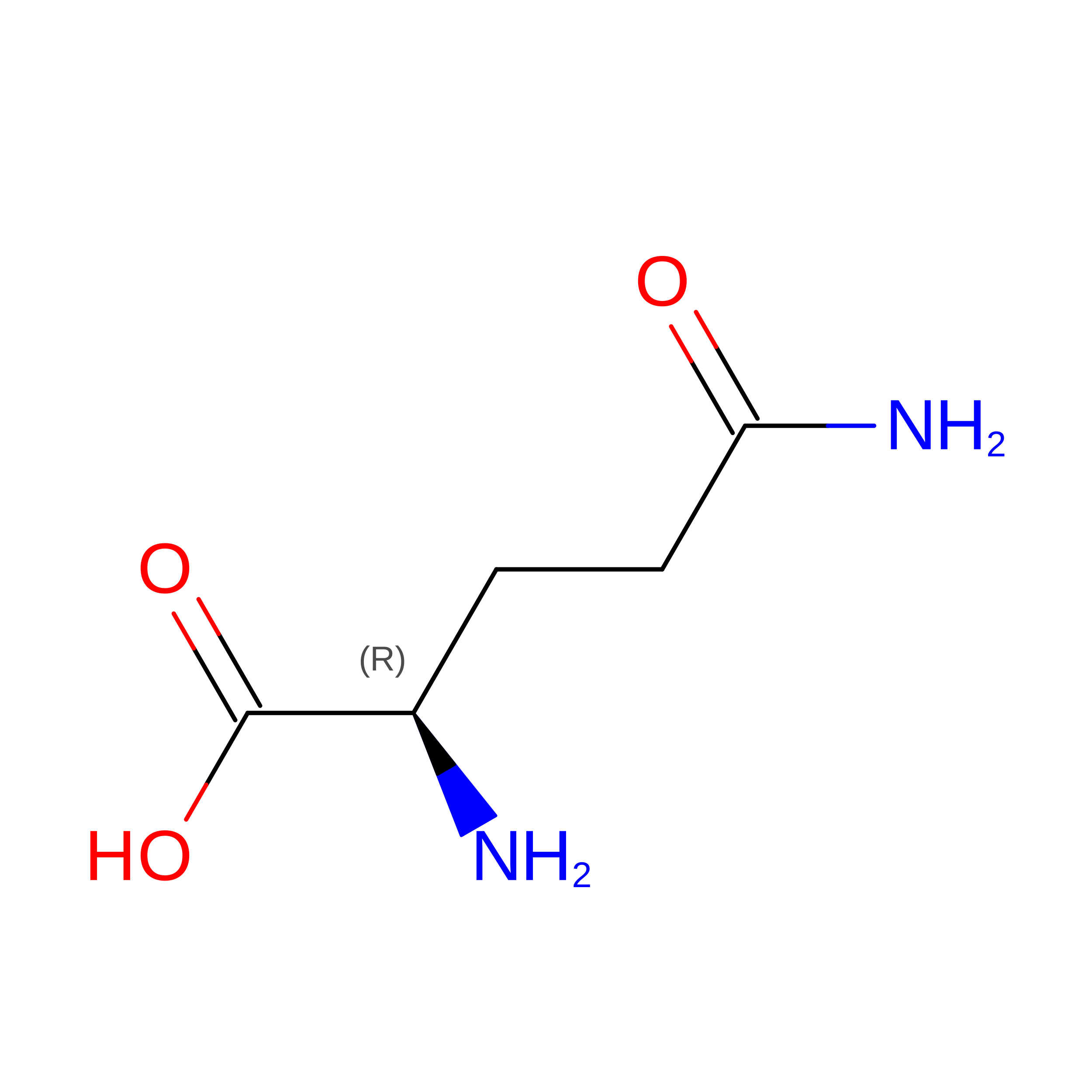
D-PEPTIDE LINKINGC3 H7 N O2

--

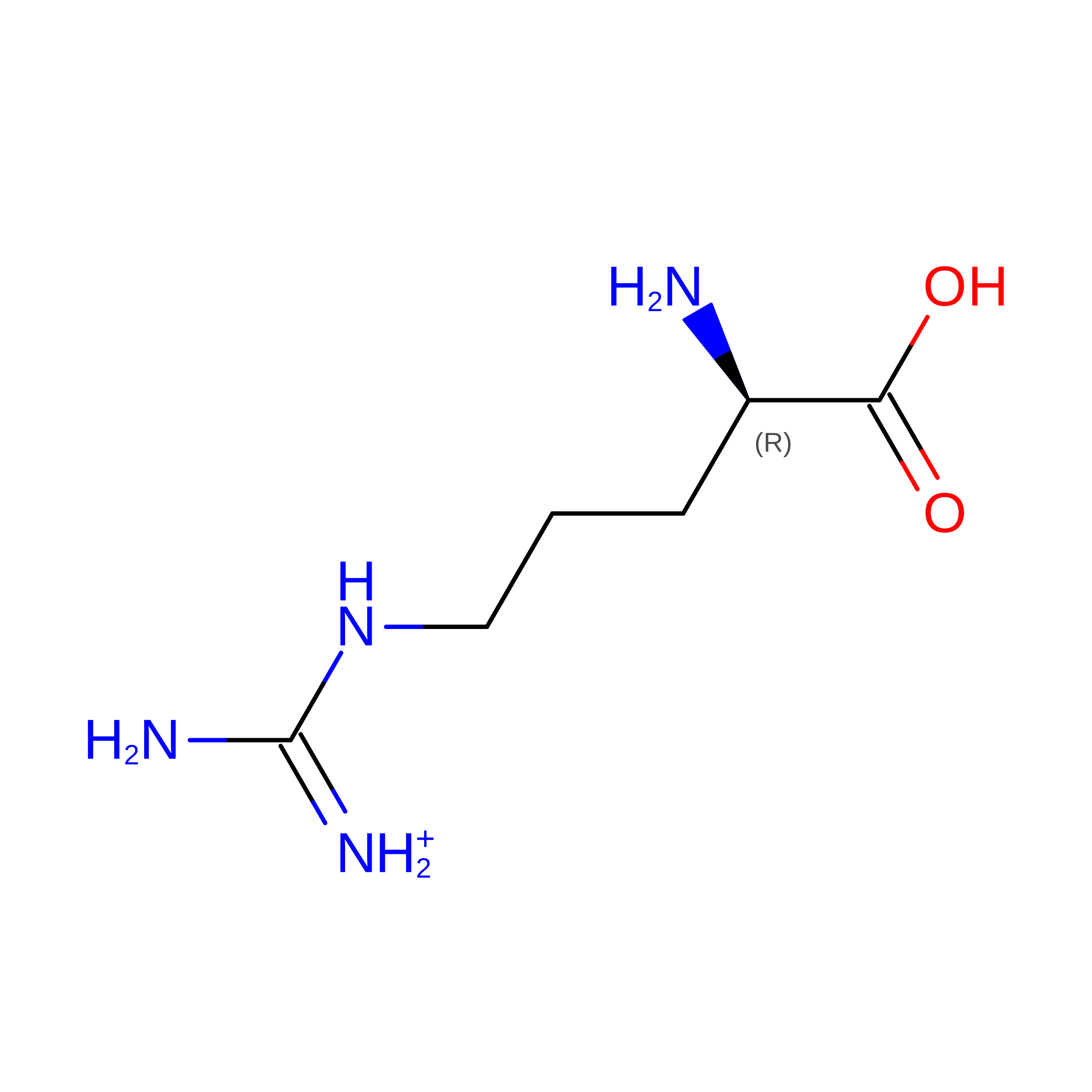
DAR
Query on DAR
A [auth D],
D [auth C]
D-PEPTIDE LINKINGC6 H15 N4 O2

--

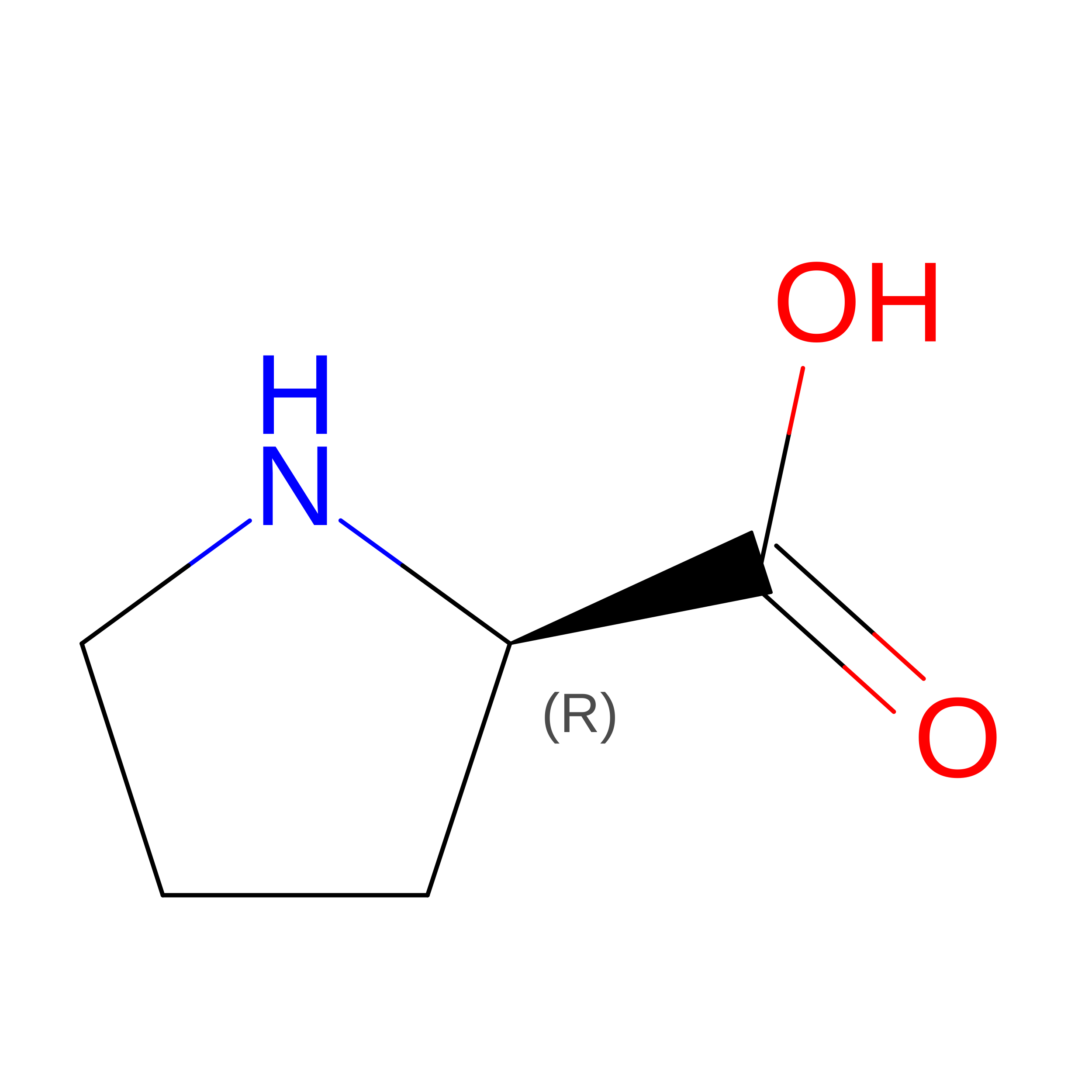
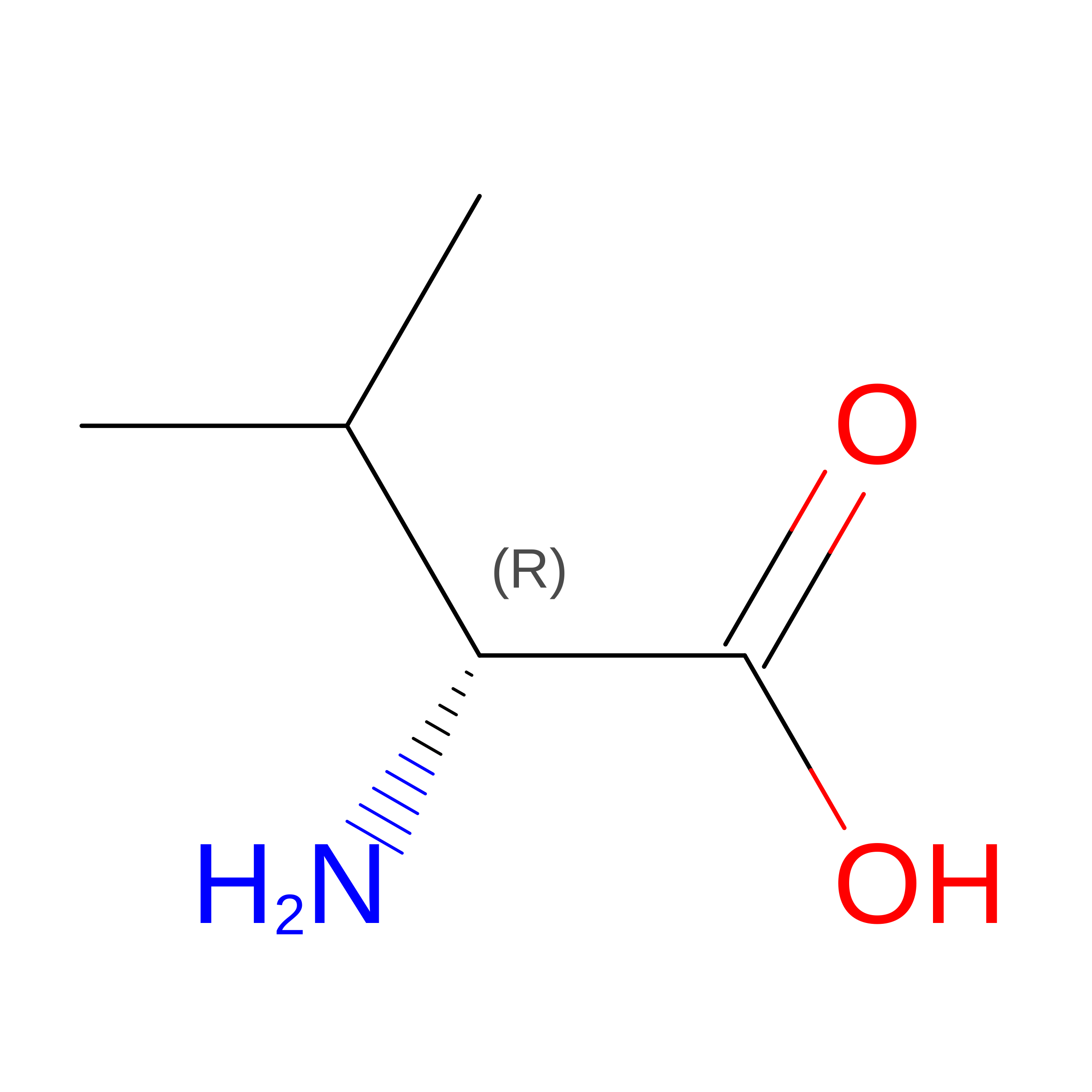
DGN
Query on DGN
A [auth D],
D [auth C]
D-PEPTIDE LINKINGC5 H10 N2 O3

--

DIL
Query on DIL
A [auth D],
D [auth C]
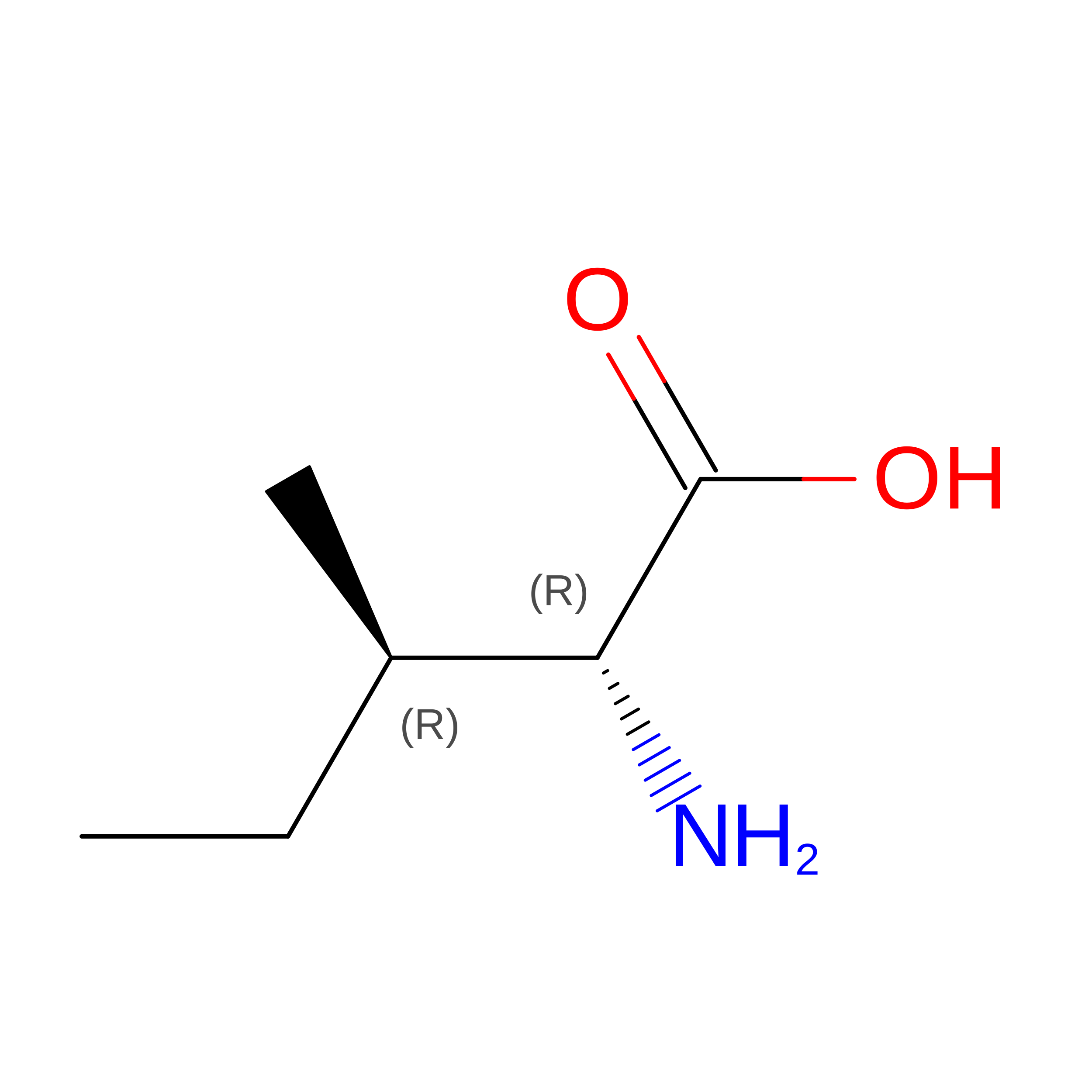
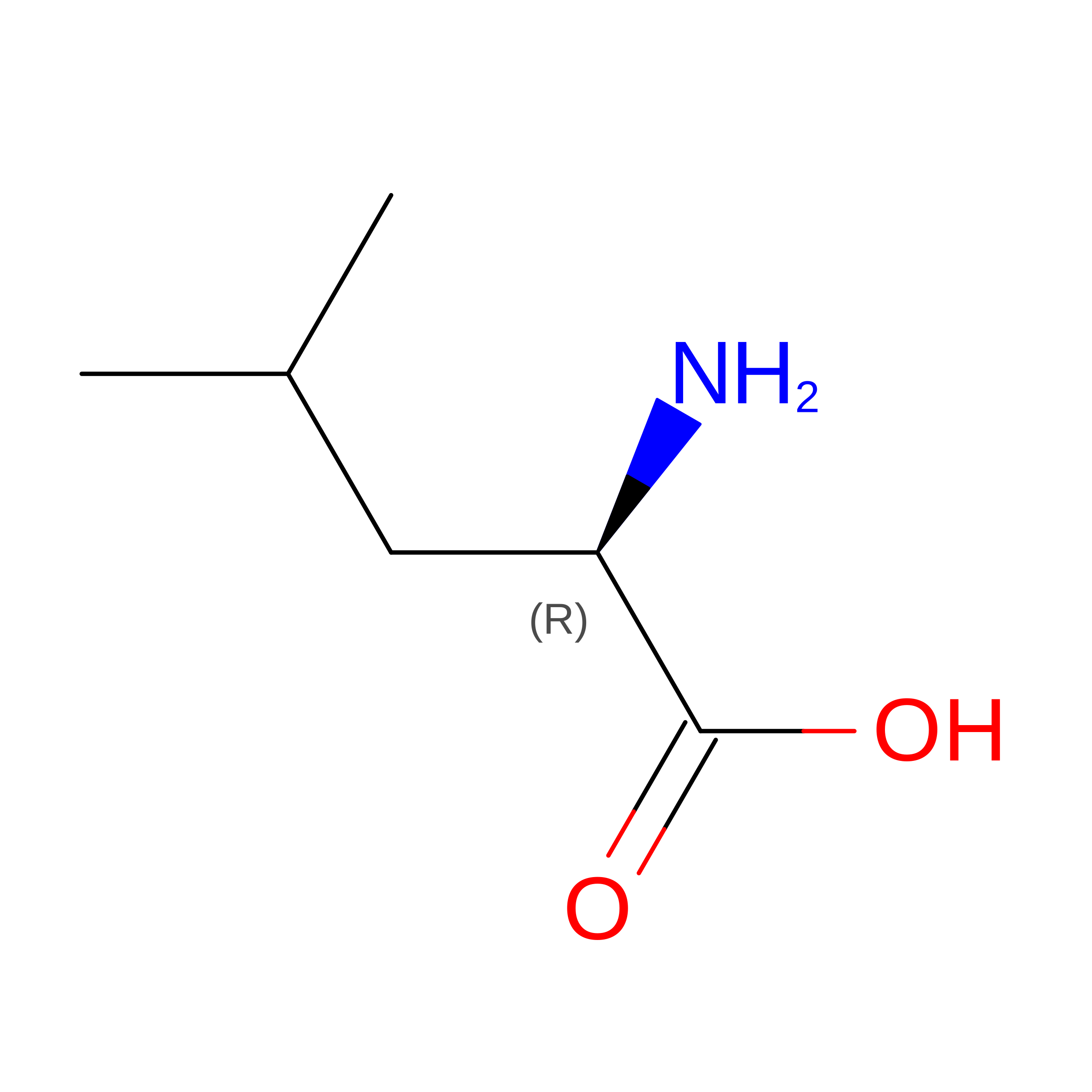
D-PEPTIDE LINKINGC6 H13 N O2

--

DLE
Query on DLE
A [auth D],
D [auth C]
D-PEPTIDE LINKINGC6 H13 N O2

--

DLY
Query on DLY
A [auth D],
D [auth C]
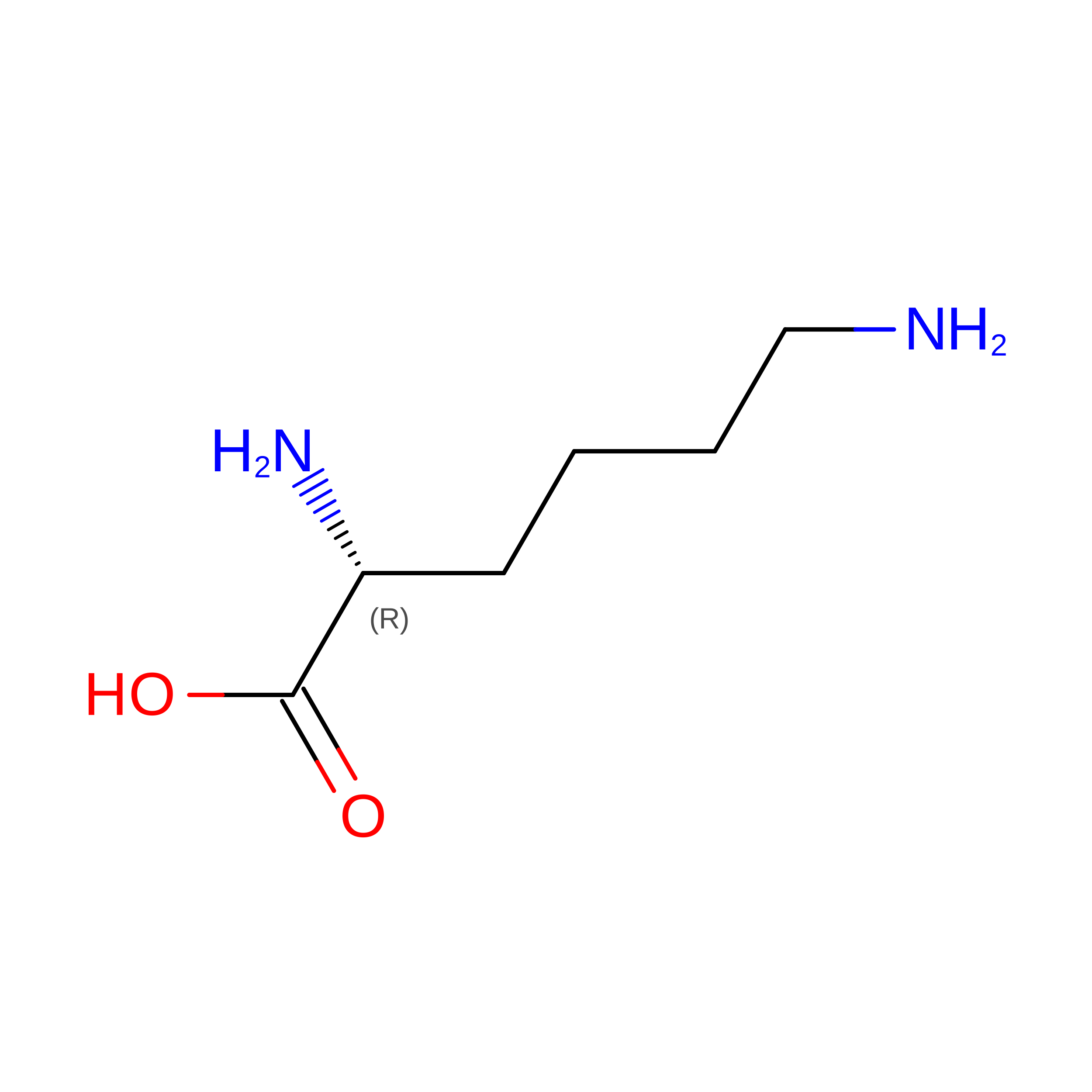
D-PEPTIDE LINKINGC6 H14 N2 O2

--

DPR
Query on DPR
A [auth D],
D [auth C]
D-PEPTIDE LINKINGC5 H9 N O2

--

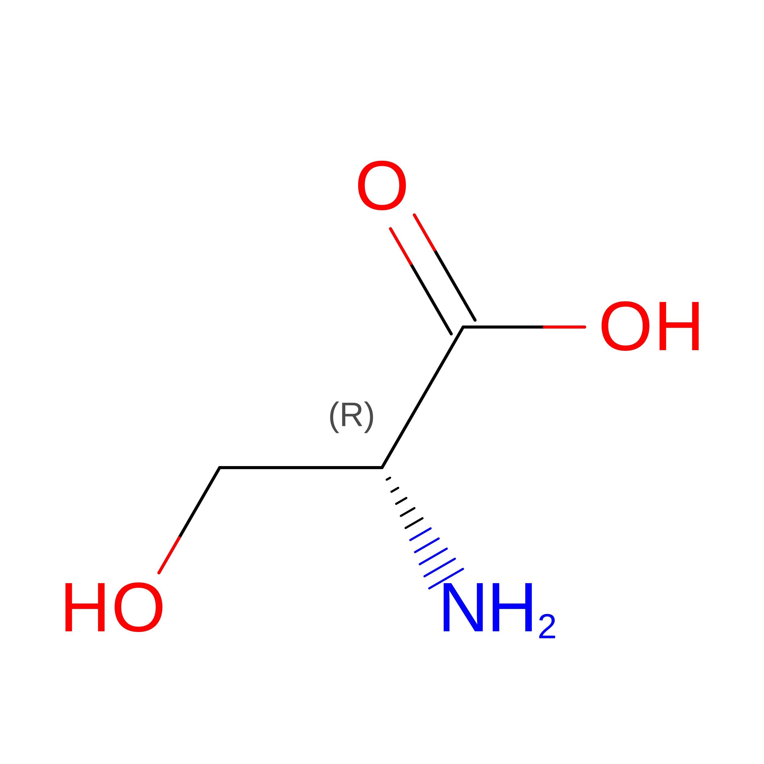
DSN
Query on DSN
A [auth D],
D [auth C]
D-PEPTIDE LINKINGC3 H7 N O3

--

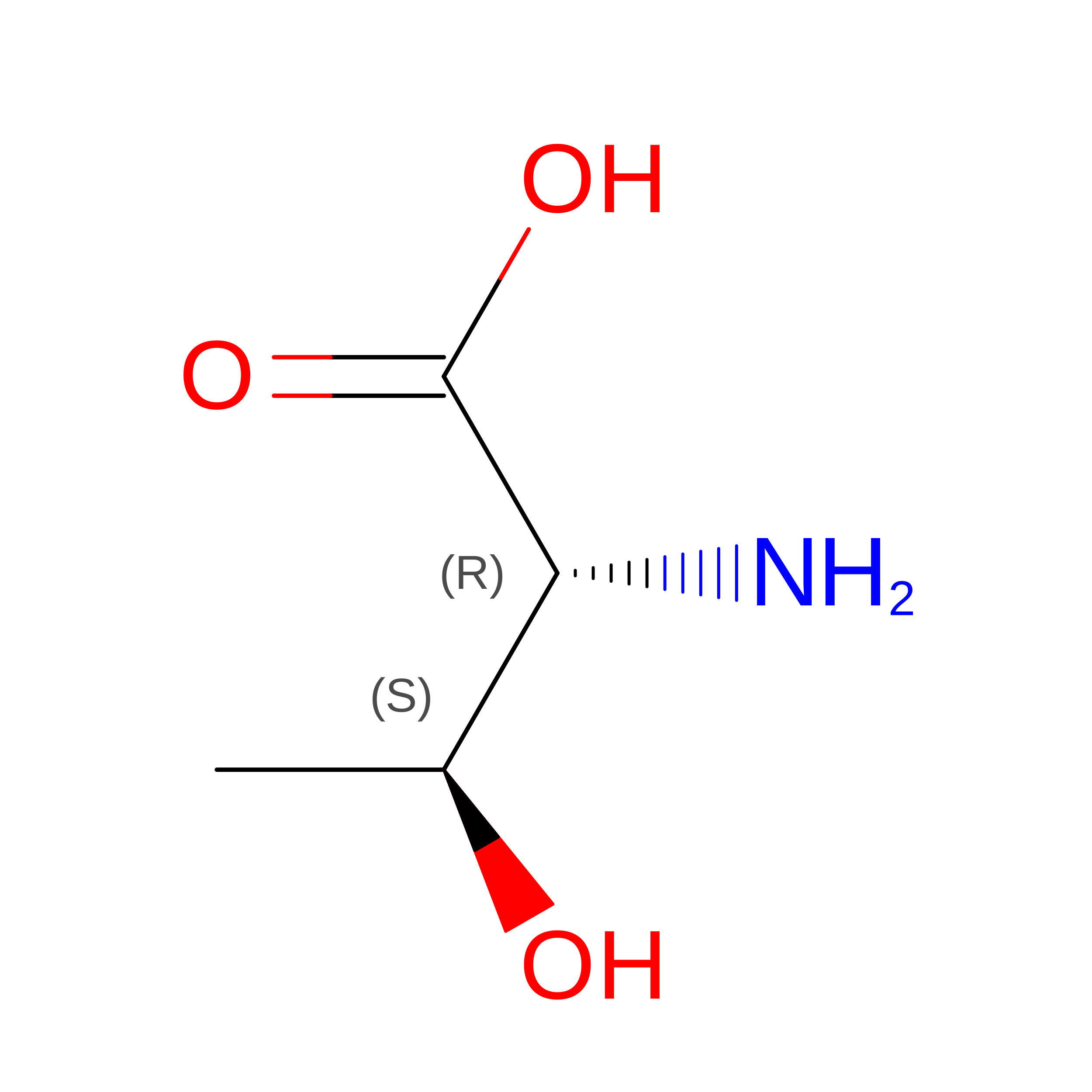
DTH
Query on DTH
A [auth D],
D [auth C]
D-PEPTIDE LINKINGC4 H9 N O3

--

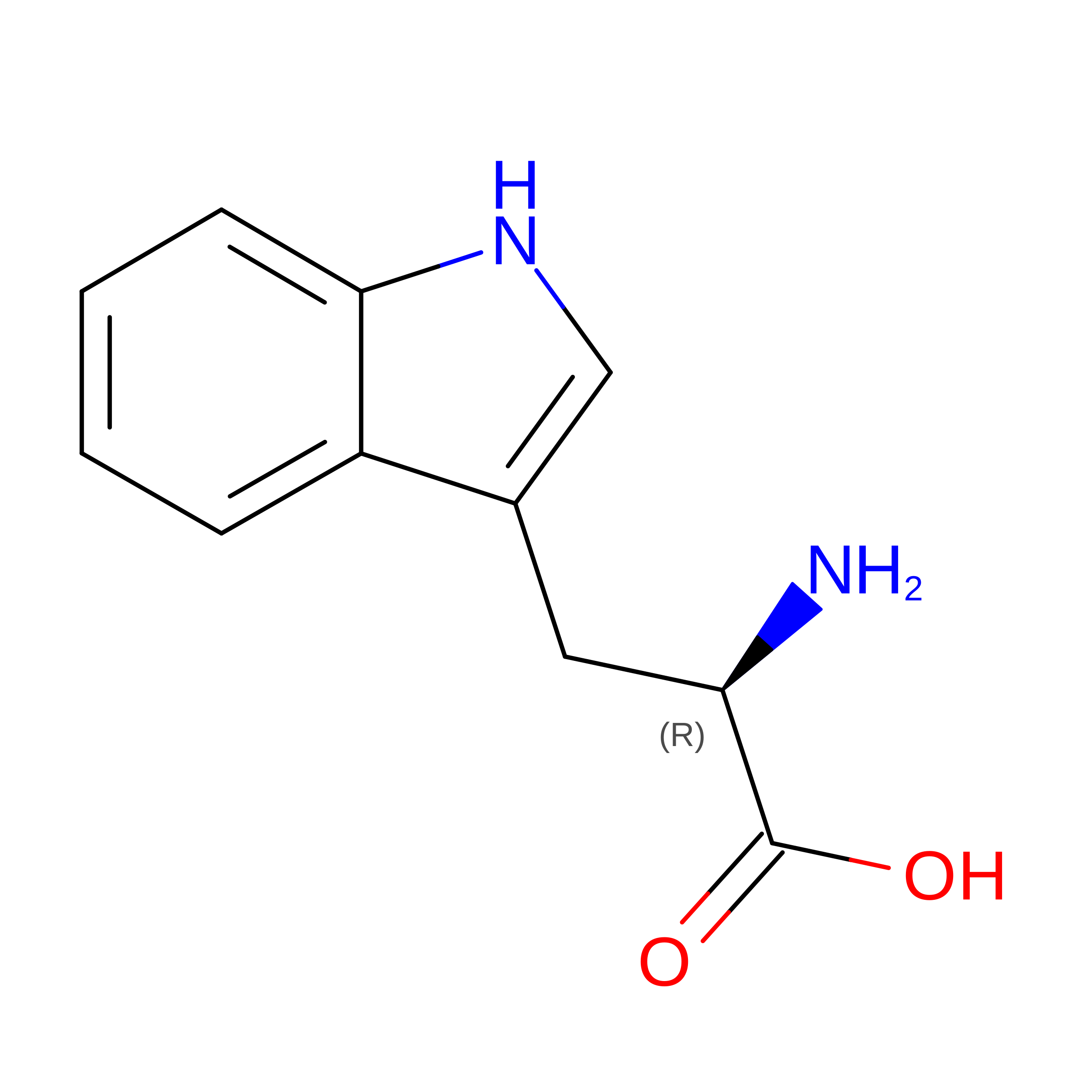
DTR
Query on DTR
A [auth D],
D [auth C]
D-PEPTIDE LINKINGC11 H12 N2 O2

--

DVA
Query on DVA
A [auth D],
D [auth C]
D-PEPTIDE LINKINGC5 H11 N O2

--

Experimental Data & Validation

Experimental Data

  • Method: X-RAY DIFFRACTION
  • Resolution: 1.27 Å
  • R-Value Free:  0.235 (Depositor), 0.235 (DCC) 
  • R-Value Work:  0.201 (Depositor), 0.200 (DCC) 
Space Group: C 1 2 1
Unit Cell:
Length ( Å )Angle ( ˚ )
a = 38.38α = 90
b = 58.99β = 110.6
c = 44.02γ = 90
Software Package:
Software NamePurpose
PHENIXrefinement
XDSdata reduction
XSCALEdata scaling
PHASERphasing

Structure Validation

View Full Validation Report


Entry History 

& Funding Information

Deposition Data


Funding OrganizationLocationGrant Number
National Institutes of Health/National Human Genome Research Institute (NIH/NHGRI)United StatesR01GM061238

Revision History  (Full details and data files)

  • Version 1.0: 2019-05-22
    Type: Initial release
  • Version 1.1: 2019-05-29
    Changes: Data collection, Database references
  • Version 1.2: 2019-12-18
    Changes: Author supporting evidence
  • Version 1.3: 2023-10-11
    Changes: Data collection, Database references, Refinement description
  • Version 1.4: 2023-11-15
    Changes: Data collection
  • Version 1.5: 2024-11-06
    Changes: Structure summary