6O38

Structure of a chaperone-substrate complex


Experimental Data Snapshot

  • Method: X-RAY DIFFRACTION
  • Resolution: 2.60 Å
  • R-Value Free: 0.240 
  • R-Value Work: 0.225 
  • R-Value Observed: 0.226 

wwPDB Validation   3D Report Full Report


This is version 1.2 of the entry. See complete history


Literature

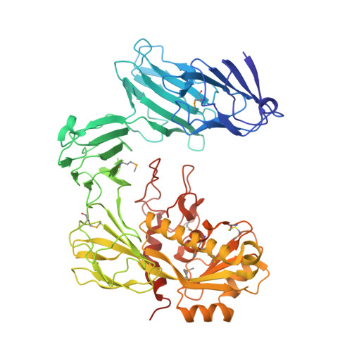
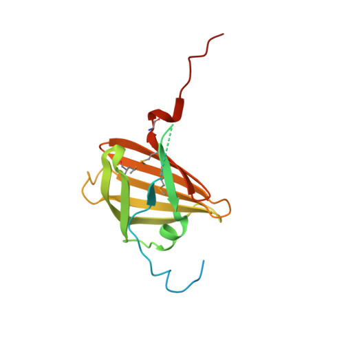
The structure ofAcinetobacter-secreted protease CpaA complexed with its chaperone CpaB reveals a novel mode of a T2SS chaperone-substrate interaction.

Urusova, D.V.Kinsella, R.L.Salinas, N.D.Haurat, M.F.Feldman, M.F.Tolia, N.H.

(2019) J Biol Chem 294: 13344-13354

  • DOI: https://doi.org/10.1074/jbc.RA119.009805
  • Primary Citation of Related Structures:  
    6O38

  • PubMed Abstract: 

    Members of the Acinetobacter baumannii-calcoaceticus complex are nosocomial pathogens frequently causing multidrug-resistant infections that are increasing at alarming rates. A. baumannii has become the Gram-negative bacterium with the highest rate of multidrug resistance. As such, it is categorized by the World Health Organization as a critical priority for the research and development of new antimicrobial therapies. The zinc-dependent metalloendopeptidase CpaA is a predominant substrate of the type II secretion system (T2SS). CpaA is also a virulence factor of medically relevant Acinetobacter strains that specifically degrade the human glycoprotein coagulation factor XII and not its deglycosylated form, but the mechanism for this specificity is unknown. CpaB is a membrane-anchored T2SS chaperone that interacts with CpaA and is required for its stability and secretion. Here, we report the crystal structure of the CpaAB complex at 2.6-Å resolution, revealing four glycan-binding domains in CpaA that were not predicted from its primary sequence and may explain CpaA's glycoprotein-targeting activity. The structure of the complex identified a novel mode for chaperone-protease interactions in which the protease surrounds the chaperone. The CpaAB organization was akin to zymogen inactivation, with CpaB serving as a prodomain that inhibits catalytically active CpaA. CpaB contains a C-terminal tail that appears to block access to the CpaA catalytic site, and functional experiments with truncated variants indicated that this tail is dispensable for CpaA expression and secretion. Our results provide new insight into the mechanism of CpaA secretion and may inform the future development of therapeutic strategies for managing Acinetobacter infections.


  • Organizational Affiliation

    Department of Molecular Microbiology, Washington University School of Medicine, Campus Box 8230, Saint Louis, Missouri 63110.


Macromolecules
Find similar proteins by:  (by identity cutoff)  |  3D Structure
Entity ID: 1
MoleculeChains Sequence LengthOrganismDetailsImage
Acinetobacter secreted protease CpaA
A, B, C, D, E
A, B, C, D, E, F
578Acinetobacter nosocomialis M2Mutation(s): 0 
UniProt
Find proteins for A0A5H1ZR49 (Acinetobacter nosocomialis M2)
Explore A0A5H1ZR49 
Go to UniProtKB:  A0A5H1ZR49
Entity Groups  
Sequence Clusters30% Identity50% Identity70% Identity90% Identity95% Identity100% Identity
UniProt GroupA0A5H1ZR49
Sequence Annotations
Expand
  • Reference Sequence
Find similar proteins by:  (by identity cutoff)  |  3D Structure
Entity ID: 2
MoleculeChains Sequence LengthOrganismDetailsImage
Type II secretion chaperone CpaB178Acinetobacter nosocomialis M2Mutation(s): 0 
Entity Groups  
Sequence Clusters30% Identity50% Identity70% Identity90% Identity95% Identity100% Identity
Sequence Annotations
Expand
  • Reference Sequence
Small Molecules
Ligands 2 Unique
IDChains Name / Formula / InChI Key2D Diagram3D Interactions
SO4
Query on SO4

Download Ideal Coordinates CCD File 
BA [auth E]
CA [auth E]
EA [auth F]
FA [auth F]
GA [auth F]
BA [auth E],
CA [auth E],
EA [auth F],
FA [auth F],
GA [auth F],
HA [auth G],
IA [auth H],
JA [auth L],
KA [auth L],
LA [auth M],
MA [auth M],
N [auth A],
NA [auth N],
O [auth A],
P [auth A],
R [auth B],
S [auth B],
T [auth B],
V [auth C],
W [auth C],
X [auth C],
Z [auth D]
SULFATE ION
O4 S
QAOWNCQODCNURD-UHFFFAOYSA-L
ZN
Query on ZN

Download Ideal Coordinates CCD File 
AA [auth E]
DA [auth F]
M [auth A]
Q [auth B]
U [auth C]
AA [auth E],
DA [auth F],
M [auth A],
Q [auth B],
U [auth C],
Y [auth D]
ZINC ION
Zn
PTFCDOFLOPIGGS-UHFFFAOYSA-N
Modified Residues  1 Unique
IDChains TypeFormula2D DiagramParent
MSE
Query on MSE
A, B, C, D, E
A, B, C, D, E, F
L-PEPTIDE LINKINGC5 H11 N O2 SeMET
Experimental Data & Validation

Experimental Data

  • Method: X-RAY DIFFRACTION
  • Resolution: 2.60 Å
  • R-Value Free: 0.240 
  • R-Value Work: 0.225 
  • R-Value Observed: 0.226 
  • Space Group: P 1
Unit Cell:
Length ( Å )Angle ( ˚ )
a = 110.747α = 108.94
b = 118.489β = 103.97
c = 129.583γ = 100.1
Software Package:
Software NamePurpose
PHENIXrefinement
HKL-3000data reduction
HKL-3000data scaling
MLPHAREphasing

Structure Validation

View Full Validation Report


Entry History 

Deposition Data

Revision History  (Full details and data files)

  • Version 1.0: 2019-07-24
    Type: Initial release
  • Version 1.1: 2019-07-31
    Changes: Data collection, Database references
  • Version 1.2: 2019-09-25
    Changes: Data collection, Database references