6O2V | pdb_00006o2v

Crystal structure of the SARAF luminal domain Cys-lock mutant monomer

  • Classification: SIGNALING PROTEIN
  • Organism(s): Homo sapiens
  • Expression System: Escherichia coli
  • Mutation(s): Yes 

  • Deposited: 2019-02-24 Released: 2019-05-29 
  • Deposition Author(s): Kimberlin, C.R., Minor, D.L.
  • Funding Organization(s): National Institutes of Health/National Heart, Lung, and Blood Institute (NIH/NHLBI), National Institutes of Health/National Institute on Deafness and Other Communication Disorders (NIH/NIDCD), American Heart Association

Experimental Data Snapshot

  • Method: X-RAY DIFFRACTION
  • Resolution: 1.58 Å
  • R-Value Free: 
    0.203 (Depositor), 0.203 (DCC) 
  • R-Value Work: 
    0.165 (Depositor), 0.167 (DCC) 
  • R-Value Observed: 
    0.167 (Depositor) 

Starting Model: experimental
View more details

wwPDB Validation 3D Report Full Report

Validation slider image for 6O2V

This is version 1.5 of the entry. See complete history

Literature

SARAF Luminal Domain Structure Reveals a Novel Domain-Swapped beta-Sandwich Fold Important for SOCE Modulation.

Kimberlin, C.R.Meshcheriakova, A.Palty, R.Raveh, A.Karbat, I.Reuveny, E.Minor Jr., D.L.

(2019) J Mol Biology 431: 2869-2883

  • DOI: https://doi.org/10.1016/j.jmb.2019.05.008
  • Primary Citation Related Structures: 
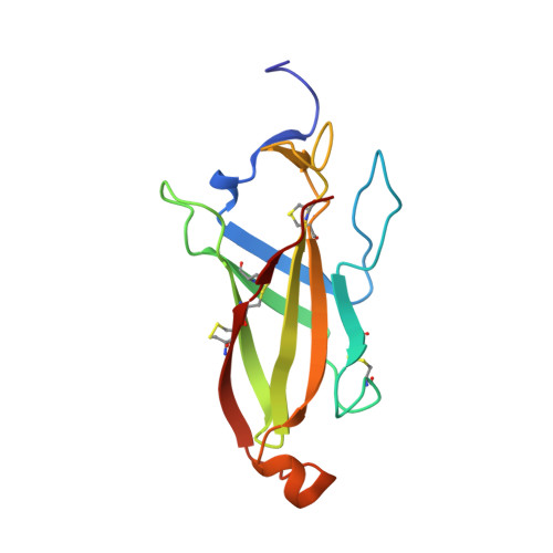
    6O2U, 6O2V, 6O2W

  • PubMed Abstract: 

    Store-Operated Calcium Entry (SOCE) plays key roles in cell proliferation, muscle contraction, immune responses, and memory formation. The coordinated interactions of a number of proteins from the plasma and endoplasmic reticulum membranes control SOCE to replenish internal Ca 2+ stores and generate intracellular Ca 2+ signals. SARAF, an endoplasmic reticulum resident component of the SOCE pathway having no homology to any characterized protein, serves as an important brake on SOCE. Here, we describe the X-ray crystal structure of the SARAF luminal domain, SARAF L . This domain forms a novel 10-stranded β-sandwich fold that includes a set of three conserved disulfide bonds, denoted the "SARAF-fold." The structure reveals a domain-swapped dimer in which the last two β-strands (β9 and β10) are exchanged forming a region denoted the "SARAF luminal switch" that is essential for dimerization. Sequence comparisons reveal that the SARAF-fold is highly conserved in vertebrates and in a variety of pathologic fungi. Förster resonance energy transfer experiments using full-length SARAF validate the formation of the domain-swapped dimer in cells and demonstrate that dimerization is reversible. A designed variant lacking the SARAF luminal switch shows that the domain swapping is essential to function and indicates that the SARAF dimer accelerates SOCE inactivation.


  • Organizational Affiliation
    • Cardiovascular Research Institute, University of California, San Francisco, CA 94148, USA.

Macromolecule Content 

  • Total Structure Weight: 31.18 kDa 
  • Atom Count: 2,505 
  • Modeled Residue Count: 270 
  • Deposited Residue Count: 270 
  • Unique protein chains: 1

Macromolecules

Find similar proteins by:|  3D Structure
Entity ID: 1
MoleculeChains  Sequence LengthOrganismDetailsImage
Store-operated calcium entry-associated regulatory factor
A, B
135Homo sapiensMutation(s): 2 
Gene Names: SARAFTMEM66XTP3HSPC035NPD003PSEC0019UNQ1967/PRO4499
UniProt & NIH Common Fund Data Resources
Find proteins for Q96BY9 (Homo sapiens)
Explore Q96BY9 
Go to UniProtKB:  Q96BY9
PHAROS:  Q96BY9
GTEx:  ENSG00000133872 
Entity Groups
Sequence Clusters30% Identity50% Identity70% Identity90% Identity95% Identity100% Identity
UniProt GroupQ96BY9
Sequence Annotations
Expand
Reference Sequence

Experimental Data & Validation

Experimental Data

  • Method: X-RAY DIFFRACTION
  • Resolution: 1.58 Å
  • R-Value Free:  0.203 (Depositor), 0.203 (DCC) 
  • R-Value Work:  0.165 (Depositor), 0.167 (DCC) 
  • R-Value Observed: 0.167 (Depositor) 
Space Group: P 21 21 21
Unit Cell:
Length ( Å )Angle ( ˚ )
a = 59.689α = 90
b = 60.953β = 90
c = 63.441γ = 90
Software Package:
Software NamePurpose
XDSdata reduction
SCALAdata scaling
PHENIXrefinement
PDB_EXTRACTdata extraction
PHASERphasing

Structure Validation

View Full Validation Report


Entry History 

& Funding Information

Deposition Data


Funding OrganizationLocationGrant Number
National Institutes of Health/National Heart, Lung, and Blood Institute (NIH/NHLBI)United StatesR01-HL080050
National Institutes of Health/National Institute on Deafness and Other Communication Disorders (NIH/NIDCD)United StatesR01-DC007664
National Institutes of Health/National Heart, Lung, and Blood Institute (NIH/NHLBI)United StatesT32HL007731
American Heart AssociationUnited States14POST18740062

Revision History  (Full details and data files)

  • Version 1.0: 2019-05-29
    Type: Initial release
  • Version 1.1: 2019-06-19
    Changes: Data collection, Database references
  • Version 1.2: 2019-07-10
    Changes: Data collection, Database references
  • Version 1.3: 2019-12-04
    Changes: Author supporting evidence
  • Version 1.4: 2023-10-11
    Changes: Data collection, Database references, Refinement description
  • Version 1.5: 2024-10-09
    Changes: Structure summary