6O2D | pdb_00006o2d

Schizosaccharomyces pombe Cnp3 Cupin Domain

  • Classification: CELL CYCLE
  • Organism(s): Schizosaccharomyces pombe
  • Expression System: Escherichia coli
  • Mutation(s): No 

  • Deposited: 2019-02-22 Released: 2019-08-21 
  • Deposition Author(s): Chik, J.K., Cho, U.S.
  • Funding Organization(s): National Institutes of Health/National Institute of Diabetes and Digestive and Kidney Disease (NIH/NIDDK), National Institutes of Health/National Institute of General Medical Sciences (NIH/NIGMS)

Experimental Data Snapshot

  • Method: X-RAY DIFFRACTION
  • Resolution: 2.52 Å
  • R-Value Free: 
    0.251 (Depositor), 0.252 (DCC) 
  • R-Value Work: 
    0.217 (Depositor), 0.220 (DCC) 
  • R-Value Observed: 
    0.219 (Depositor) 

wwPDB Validation 3D Report Full Report

Validation slider image for 6O2D

This is version 1.4 of the entry. See complete history

Literature

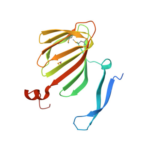
Structures of CENP-C cupin domains at regional centromeres reveal unique patterns of dimerization and recruitment functions for the inner pocket.

Chik, J.K.Moiseeva, V.Goel, P.K.Meinen, B.A.Koldewey, P.An, S.Mellone, B.G.Subramanian, L.Cho, U.S.

(2019) J Biological Chem 294: 14119-14134

  • DOI: https://doi.org/10.1074/jbc.RA119.008464
  • Primary Citation Related Structures: 
    6O2D, 6O2K

  • PubMed Abstract: 

    The successful assembly and regulation of the kinetochore are critical for the equal and accurate segregation of genetic material during the cell cycle. CENP-C (centromere protein C), a conserved inner kinetochore component, has been broadly characterized as a scaffolding protein and is required for the recruitment of multiple kinetochore proteins to the centromere. At its C terminus, CENP-C harbors a conserved cupin domain that has an established role in protein dimerization. Although the crystal structure of the Saccharomyces cerevisiae Mif2 CENP-C cupin domain has been determined, centromeric organization and kinetochore composition vary greatly between S. cerevisiae (point centromere) and other eukaryotes (regional centromere). Therefore, whether the structural and functional role of the cupin domain is conserved throughout evolution requires investigation. Here, we report the crystal structures of the Schizosaccharomyces pombe and Drosophila melanogaster CENP-C cupin domains at 2.52 and 1.81 Å resolutions, respectively. Although the central jelly roll architecture is conserved among the three determined CENP-C cupin domain structures, the cupin domains from organisms with regional centromeres contain additional structural features that aid in dimerization. Moreover, we found that the S. pombe Cnp3 CENP-C jelly roll fold harbors an inner binding pocket that is used to recruit the meiosis-specific protein Moa1. In summary, our results unveil the evolutionarily conserved and unique features of the CENP-C cupin domain and uncover the mechanism by which it functions as a recruitment factor.


  • Organizational Affiliation
    • Department of Biological Chemistry, University of Michigan Medical School, Ann Arbor, Michigan 48109.

Macromolecule Content 

  • Total Structure Weight: 36.1 kDa 
  • Atom Count: 2,251 
  • Modeled Residue Count: 282 
  • Deposited Residue Count: 316 
  • Unique protein chains: 1

Macromolecules

Find similar proteins by:|  3D Structure
Entity ID: 1
MoleculeChains  Sequence LengthOrganismDetailsImage
Inner kinetochore subunit cnp3
A, B
158Schizosaccharomyces pombeMutation(s): 0 
Gene Names: cnp3SPBC1861.01cSPBC56F2.13
UniProt
Find proteins for Q9USR9 (Schizosaccharomyces pombe (strain 972 / ATCC 24843))
Explore Q9USR9 
Go to UniProtKB:  Q9USR9
Entity Groups
Sequence Clusters30% Identity50% Identity70% Identity90% Identity95% Identity100% Identity
UniProt GroupQ9USR9
Sequence Annotations
Expand
Reference Sequence

Small Molecules

Modified Residues  1 Unique
IDChains TypeFormula2D DiagramParent
MSE
Query on MSE
A, B
L-PEPTIDE LINKINGC5 H11 N O2 SeMET

Experimental Data & Validation

Experimental Data

  • Method: X-RAY DIFFRACTION
  • Resolution: 2.52 Å
  • R-Value Free:  0.251 (Depositor), 0.252 (DCC) 
  • R-Value Work:  0.217 (Depositor), 0.220 (DCC) 
  • R-Value Observed: 0.219 (Depositor) 
Space Group: P 41 21 2
Unit Cell:
Length ( Å )Angle ( ˚ )
a = 55.16α = 90
b = 55.16β = 90
c = 206.64γ = 90
Software Package:
Software NamePurpose
PHENIXrefinement
Cootmodel building
AutoSolphasing
XDSdata scaling
XDSdata reduction

Structure Validation

View Full Validation Report


Entry History 

& Funding Information

Deposition Data


Funding OrganizationLocationGrant Number
National Institutes of Health/National Institute of Diabetes and Digestive and Kidney Disease (NIH/NIDDK)United StatesR01-DK111465
National Institutes of Health/National Institute of General Medical Sciences (NIH/NIGMS)United StatesT32-GM007315

Revision History  (Full details and data files)

  • Version 1.0: 2019-08-21
    Type: Initial release
  • Version 1.1: 2019-09-25
    Changes: Data collection, Database references
  • Version 1.2: 2019-10-02
    Changes: Data collection, Database references
  • Version 1.3: 2019-12-25
    Changes: Author supporting evidence
  • Version 1.4: 2024-10-23
    Changes: Data collection, Database references, Structure summary