6O1V

Complex of human cystic fibrosis transmembrane conductance regulator (CFTR) and GLPG1837


Experimental Data Snapshot

  • Method: ELECTRON MICROSCOPY
  • Resolution: 3.20 Å
  • Aggregation State: PARTICLE 
  • Reconstruction Method: SINGLE PARTICLE 

wwPDB Validation   3D Report Full Report


This is version 1.3 of the entry. See complete history


Literature

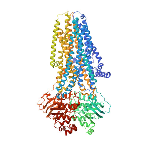
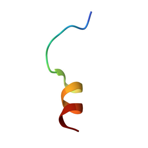
Structural identification of a hotspot on CFTR for potentiation.

Liu, F.Zhang, Z.Levit, A.Levring, J.Touhara, K.K.Shoichet, B.K.Chen, J.

(2019) Science 364: 1184-1188

  • DOI: https://doi.org/10.1126/science.aaw7611
  • Primary Citation of Related Structures:  
    6O1V, 6O2P

  • PubMed Abstract: 

    Cystic fibrosis is a fatal disease caused by mutations in the cystic fibrosis transmembrane conductance regulator (CFTR). Two main categories of drugs are being developed: correctors that improve folding of CFTR and potentiators that recover the function of CFTR. Here, we report two cryo-electron microscopy structures of human CFTR in complex with potentiators: one with the U.S. Food and Drug Administration (FDA)-approved drug ivacaftor at 3.3-angstrom resolution and the other with an investigational drug, GLPG1837, at 3.2-angstrom resolution. These two drugs, although chemically dissimilar, bind to the same site within the transmembrane region. Mutagenesis suggests that in both cases, hydrogen bonds provided by the protein are important for drug recognition. The molecular details of how ivacaftor and GLPG1837 interact with CFTR may facilitate structure-based optimization of therapeutic compounds.


  • Organizational Affiliation

    Laboratory of Membrane Biophysics and Biology, The Rockefeller University, New York, NY 10065, USA.


Macromolecules
Find similar proteins by:  (by identity cutoff)  |  3D Structure
Entity ID: 1
MoleculeChains Sequence LengthOrganismDetailsImage
Cystic fibrosis transmembrane conductance regulator1,489Homo sapiensMutation(s): 1 
Gene Names: CFTRABCC7
EC: 3.6.3.49
Membrane Entity: Yes 
UniProt & NIH Common Fund Data Resources
Find proteins for P13569 (Homo sapiens)
Explore P13569 
Go to UniProtKB:  P13569
PHAROS:  P13569
GTEx:  ENSG00000001626 
Entity Groups  
Sequence Clusters30% Identity50% Identity70% Identity90% Identity95% Identity100% Identity
UniProt GroupP13569
Sequence Annotations
Expand
  • Reference Sequence

Find similar proteins by:  Sequence   |   3D Structure  

Entity ID: 2
MoleculeChains Sequence LengthOrganismDetailsImage
Unknown Peptide17Homo sapiensMutation(s): 0 
Sequence Annotations
Expand
  • Reference Sequence
Small Molecules
Ligands 5 Unique
IDChains Name / Formula / InChI Key2D Diagram3D Interactions
POV
Query on POV

Download Ideal Coordinates CCD File 
H [auth A],
I [auth A],
J [auth A],
K [auth A],
L [auth A]
(2S)-3-(hexadecanoyloxy)-2-[(9Z)-octadec-9-enoyloxy]propyl 2-(trimethylammonio)ethyl phosphate
C42 H82 N O8 P
WTJKGGKOPKCXLL-PFDVCBLKSA-N
ATP
Query on ATP

Download Ideal Coordinates CCD File 
F [auth A],
G [auth A]
ADENOSINE-5'-TRIPHOSPHATE
C10 H16 N5 O13 P3
ZKHQWZAMYRWXGA-KQYNXXCUSA-N
CLR
Query on CLR

Download Ideal Coordinates CCD File 
M [auth A]CHOLESTEROL
C27 H46 O
HVYWMOMLDIMFJA-DPAQBDIFSA-N
LJP
Query on LJP

Download Ideal Coordinates CCD File 
C [auth A]N-(3-carbamoyl-5,5,7,7-tetramethyl-4,7-dihydro-5H-thieno[2,3-c]pyran-2-yl)-1H-pyrazole-3-carboxamide
C16 H20 N4 O3 S
GHTGYZMBQPXTCQ-UHFFFAOYSA-N
MG
Query on MG

Download Ideal Coordinates CCD File 
D [auth A],
E [auth A]
MAGNESIUM ION
Mg
JLVVSXFLKOJNIY-UHFFFAOYSA-N
Experimental Data & Validation

Experimental Data

  • Method: ELECTRON MICROSCOPY
  • Resolution: 3.20 Å
  • Aggregation State: PARTICLE 
  • Reconstruction Method: SINGLE PARTICLE 
EM Software:
TaskSoftware PackageVersion
RECONSTRUCTIONFREALIGN
MODEL REFINEMENTREFMAC

Structure Validation

View Full Validation Report


Entry History & Funding Information

Deposition Data


Funding OrganizationLocationGrant Number
Howard Hughes Medical Institute (HHMI)United States--

Revision History  (Full details and data files)

  • Version 1.0: 2019-06-26
    Type: Initial release
  • Version 1.1: 2019-07-03
    Changes: Data collection, Database references
  • Version 1.2: 2019-11-20
    Changes: Author supporting evidence
  • Version 1.3: 2019-11-27
    Changes: Data collection