6NWH | pdb_00006nwh

Structures of the transcriptional regulator BgaR, a lactose sensor.


Experimental Data Snapshot

  • Method: X-RAY DIFFRACTION
  • Resolution: 2.03 Å
  • R-Value Free: 
    0.206 (Depositor), 0.218 (DCC) 
  • R-Value Work: 
    0.179 (Depositor), 0.191 (DCC) 
  • R-Value Observed: 
    0.180 (Depositor) 

wwPDB Validation 3D Report Full Report

Validation slider image for 6NWH

This is version 2.1 of the entry. See complete history

Literature

Structures of the transcriptional regulator BgaR, a lactose sensor.

Newman, J.Caron, K.Nebl, T.Peat, T.S.

(2019) Acta Crystallogr D Struct Biol 75: 639-646

  • DOI: https://doi.org/10.1107/S2059798319008131
  • Primary Citation Related Structures: 
    6NWH, 6NWJ, 6NWM, 6NWO, 6NX3

  • PubMed Abstract: 

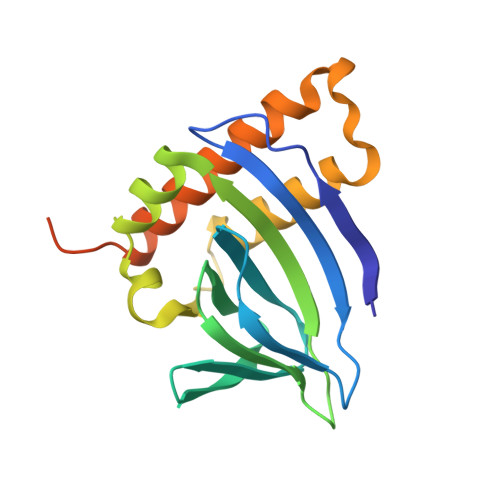
    The structure of BgaR, a transcriptional regulator of the lactose operon in Clostridium perfringens, has been solved by SAD phasing using a mercury derivative. BgaR is an exquisite sensor of lactose, with a binding affinity in the low-micromolar range. This sensor and regulator has been captured bound to lactose and to lactulose as well as in a nominal apo form, and was compared with AraC, another saccharide-binding transcriptional regulator. It is shown that the saccharides bind in the N-terminal region of a jelly-roll fold, but that part of the saccharide is exposed to bulk solvent. This differs from the classical AraC saccharide-binding site, which is mostly sequestered from the bulk solvent. The structures of BgaR bound to lactose and to lactulose highlight how specific and nonspecific interactions lead to a higher binding affinity of BgaR for lactose compared with lactulose. Moreover, solving multiple structures of BgaR in different space groups, both bound to saccharides and unbound, verified that the dimer interface along a C-terminal helix is similar to the dimer interface observed in AraC.


  • Organizational Affiliation
    • Biomedical Program, CSIRO, 343 Royal Parade, Parkville, VIC 3052, Australia.

Macromolecule Content 

  • Total Structure Weight: 45.02 kDa 
  • Atom Count: 3,003 
  • Modeled Residue Count: 325 
  • Deposited Residue Count: 364 
  • Unique protein chains: 1

Macromolecules

Find similar proteins by:|  3D Structure
Entity ID: 1
MoleculeChains  Sequence LengthOrganismDetailsImage
Transcriptional regulator BgaR
A, B
182Clostridium perfringens str. 13Mutation(s): 0 
Gene Names: CPE0770
UniProt
Find proteins for Q8XMB9 (Clostridium perfringens (strain 13 / Type A))
Explore Q8XMB9 
Go to UniProtKB:  Q8XMB9
Entity Groups
Sequence Clusters30% Identity50% Identity70% Identity90% Identity95% Identity100% Identity
UniProt GroupQ8XMB9
Sequence Annotations
Expand
Reference Sequence

Oligosaccharides

Help  
Entity ID: 2
MoleculeChains Length2D Diagram GlycosylationD Interactions
beta-D-galactopyranose-(1-4)-beta-D-glucopyranose
C, D
2N/A
Glycosylation Resources
GlyTouCan: G84224TW
GlyCosmos: G84224TW
GlyGen: G84224TW

Small Molecules

Ligands 2 Unique
IDChains Name / Formula / InChI Key2D Diagram3D Interactions
HG

Query on HG


Download:Ideal Coordinates CCD File
E [auth A]
F [auth A]
G [auth A]
H [auth A]
I [auth A]
E [auth A],
F [auth A],
G [auth A],
H [auth A],
I [auth A],
J [auth B],
K [auth B],
L [auth B],
M [auth B]
MERCURY (II) ION
Hg
BQPIGGFYSBELGY-UHFFFAOYSA-N
CL

Query on CL


Download:Ideal Coordinates CCD File
N [auth B]CHLORIDE ION
Cl
VEXZGXHMUGYJMC-UHFFFAOYSA-M

Biologically Interesting Molecules (External Reference) 

1 Unique

Experimental Data & Validation

Experimental Data

  • Method: X-RAY DIFFRACTION
  • Resolution: 2.03 Å
  • R-Value Free:  0.206 (Depositor), 0.218 (DCC) 
  • R-Value Work:  0.179 (Depositor), 0.191 (DCC) 
  • R-Value Observed: 0.180 (Depositor) 
Space Group: P 2 21 21
Unit Cell:
Length ( Å )Angle ( ˚ )
a = 46.168α = 90
b = 86.568β = 90
c = 119.812γ = 90
Software Package:
Software NamePurpose
REFMACrefinement
ADDREFdata reduction
Aimlessdata scaling
CRANK2phasing

Structure Validation

View Full Validation Report


Entry History 

Deposition Data

Revision History  (Full details and data files)

  • Version 1.0: 2019-07-17
    Type: Initial release
  • Version 1.1: 2019-07-24
    Changes: Data collection, Database references
  • Version 2.0: 2020-07-29
    Type: Remediation
    Reason: Carbohydrate remediation
    Changes: Atomic model, Author supporting evidence, Data collection, Derived calculations, Non-polymer description, Structure summary
  • Version 2.1: 2024-03-13
    Changes: Data collection, Database references, Refinement description, Structure summary