6NW9 | pdb_00006nw9

CRYSTAL STRUCTURE OF A TAILSPIKE PROTEIN 3 (TSP3, ORF212) FROM ESCHERICHIA COLI O157:H7 BACTERIOPHAGE CBA120


Experimental Data Snapshot

  • Method: X-RAY DIFFRACTION
  • Resolution: 1.85 Å
  • R-Value Free: 
    0.184 (Depositor), 0.184 (DCC) 
  • R-Value Work: 
    0.151 (Depositor), 0.151 (DCC) 
  • R-Value Observed: 
    0.152 (Depositor) 

wwPDB Validation 3D Report Full Report

Validation slider image for 6NW9

This is version 1.2 of the entry. See complete history

Literature

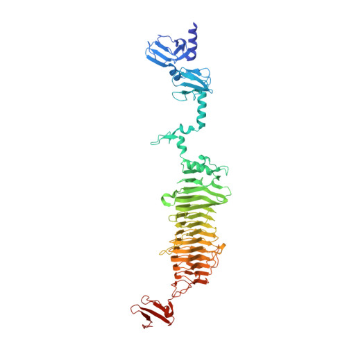
Structure and tailspike glycosidase machinery of ORF212 from E. coli O157:H7 phage CBA120 (TSP3).

Greenfield, J.Shang, X.Luo, H.Zhou, Y.Heselpoth, R.D.Nelson, D.C.Herzberg, O.

(2019) Sci Rep 9: 7349-7349

  • DOI: https://doi.org/10.1038/s41598-019-43748-9
  • Primary Citation Related Structures: 
    6NW9

  • PubMed Abstract: 

    Bacteriophage tailspike proteins mediate virion absorption through reversible primary receptor binding, followed by lipopolysaccharide or exopolysaccharide degradation. The Escherichia coli O157:H7 bacteriophage CBA120 genome encodes four distinct tailspike proteins, annotated as ORFs 210 through 213. Previously, we reported the crystal structure of ORF210 (TSP1). Here we describe the crystal structure of ORF212 (TSP3) determined at 1.85 Å resolution. As observed with other tailspike proteins, TSP3 assembles into a trimer. Each subunit of TSP3 has an N-terminal head domain that is structurally similar to that of TSP1, consistent with their high amino acid sequence identity. In contrast, despite sharing a β-helix fold, the overall structure of the C-terminal catalytic domain of TSP3 is quite different when compared to TSP1. The TSP3 structure suggests that the glycosidase active site resides in a cleft at the interface between two adjacent subunits where three acidic residues, Glu362 and Asp383 on one subunit, and Asp426 on a second subunit, are located in close proximity. Comparing the glycosidase activity of wild-type TSP3 to various point mutants revealed that catalysis requires the carboxyl groups of Glu362 and Asp426, and not of Asp383, confirming the enzyme employs two carboxyl groups to degrade lippopolysaccharide using an acid/base mechanism.


  • Organizational Affiliation
    • Institute for Bioscience and Biotechnology Research, University of Maryland, Rockville, Maryland, USA.

Macromolecule Content 

  • Total Structure Weight: 213.21 kDa 
  • Atom Count: 16,468 
  • Modeled Residue Count: 1,851 
  • Deposited Residue Count: 1,899 
  • Unique protein chains: 1

Macromolecules

Find similar proteins by:|  3D Structure
Entity ID: 1
MoleculeChains  Sequence LengthOrganismDetailsImage
Tailspike protein
A, B, C
633Kuttervirus CBA120Mutation(s): 0 
Gene Names: orf212
UniProt
Find proteins for G3M191 (Escherichia phage Cba120)
Explore G3M191 
Go to UniProtKB:  G3M191
Entity Groups
Sequence Clusters30% Identity50% Identity70% Identity90% Identity95% Identity100% Identity
UniProt GroupG3M191
Sequence Annotations
Expand
Reference Sequence

Small Molecules

Ligands 9 Unique
IDChains Name / Formula / InChI Key2D Diagram3D Interactions
PGE

Query on PGE


Download:Ideal Coordinates CCD File
FA [auth A]TRIETHYLENE GLYCOL
C6 H14 O4
ZIBGPFATKBEMQZ-UHFFFAOYSA-N
PEG

Query on PEG


Download:Ideal Coordinates CCD File
DA [auth A]
EA [auth A]
LB [auth B]
MB [auth B]
NB [auth B]
DA [auth A],
EA [auth A],
LB [auth B],
MB [auth B],
NB [auth B],
TC [auth C]
DI(HYDROXYETHYL)ETHER
C4 H10 O3
MTHSVFCYNBDYFN-UHFFFAOYSA-N
PO4

Query on PO4


Download:Ideal Coordinates CCD File
GA [auth A],
IA [auth A]
PHOSPHATE ION
O4 P
NBIIXXVUZAFLBC-UHFFFAOYSA-K
GOL

Query on GOL


Download:Ideal Coordinates CCD File
AC [auth C]
BC [auth C]
CC [auth C]
HA [auth A]
J [auth A]
AC [auth C],
BC [auth C],
CC [auth C],
HA [auth A],
J [auth A],
JA [auth A],
K [auth A],
KA [auth A],
L [auth A],
M [auth A],
N [auth A],
O [auth A],
P [auth A],
Q [auth A],
QA [auth B],
R [auth A],
RA [auth B],
S [auth A],
SA [auth B],
SB [auth C],
TA [auth B],
TB [auth C],
UA [auth B],
UB [auth C],
VA [auth B],
VB [auth C],
WA [auth B],
WB [auth C],
XA [auth B],
XB [auth C],
YA [auth B],
YB [auth C],
ZA [auth B],
ZB [auth C]
GLYCEROL
C3 H8 O3
PEDCQBHIVMGVHV-UHFFFAOYSA-N
EDO

Query on EDO


Download:Ideal Coordinates CCD File
AA [auth A]
AB [auth B]
BA [auth A]
BB [auth B]
CA [auth A]
AA [auth A],
AB [auth B],
BA [auth A],
BB [auth B],
CA [auth A],
CB [auth B],
DB [auth B],
DC [auth C],
EB [auth B],
EC [auth C],
FB [auth B],
FC [auth C],
GB [auth B],
GC [auth C],
HB [auth B],
HC [auth C],
IB [auth B],
IC [auth C],
JB [auth B],
JC [auth C],
KB [auth B],
KC [auth C],
LA [auth A],
LC [auth C],
MC [auth C],
NC [auth C],
OC [auth C],
PB [auth B],
PC [auth C],
QC [auth C],
RC [auth C],
SC [auth C],
T [auth A],
U [auth A],
V [auth A],
W [auth A],
X [auth A],
Y [auth A],
Z [auth A]
1,2-ETHANEDIOL
C2 H6 O2
LYCAIKOWRPUZTN-UHFFFAOYSA-N
CO3

Query on CO3


Download:Ideal Coordinates CCD File
H [auth A],
I [auth A],
MA [auth B]
CARBONATE ION
C O3
BVKZGUZCCUSVTD-UHFFFAOYSA-L
CL

Query on CL


Download:Ideal Coordinates CCD File
D [auth A]
NA [auth B]
OA [auth B]
OB [auth B]
QB [auth C]
D [auth A],
NA [auth B],
OA [auth B],
OB [auth B],
QB [auth C],
RB [auth C]
CHLORIDE ION
Cl
VEXZGXHMUGYJMC-UHFFFAOYSA-M
MG

Query on MG


Download:Ideal Coordinates CCD File
G [auth A]MAGNESIUM ION
Mg
JLVVSXFLKOJNIY-UHFFFAOYSA-N
NA

Query on NA


Download:Ideal Coordinates CCD File
E [auth A],
F [auth A],
PA [auth B]
SODIUM ION
Na
FKNQFGJONOIPTF-UHFFFAOYSA-N

Experimental Data & Validation

Experimental Data

  • Method: X-RAY DIFFRACTION
  • Resolution: 1.85 Å
  • R-Value Free:  0.184 (Depositor), 0.184 (DCC) 
  • R-Value Work:  0.151 (Depositor), 0.151 (DCC) 
  • R-Value Observed: 0.152 (Depositor) 
Space Group: P 1 21 1
Unit Cell:
Length ( Å )Angle ( ˚ )
a = 99.439α = 90
b = 66.77β = 103.58
c = 161.6γ = 90
Software Package:
Software NamePurpose
PHENIXrefinement
HKL-3000data reduction
HKL-3000data scaling
HKL-3000phasing

Structure Validation

View Full Validation Report


Entry History 

& Funding Information

Deposition Data


Funding OrganizationLocationGrant Number
National Institutes of Health/National Human Genome Research Institute (NIH/NHGRI)United StatesR01GM110202

Revision History  (Full details and data files)

  • Version 1.0: 2019-06-05
    Type: Initial release
  • Version 1.1: 2019-12-18
    Changes: Author supporting evidence
  • Version 1.2: 2024-03-13
    Changes: Data collection, Database references, Derived calculations