Mg2+ binds to the surface of thymidylate synthase and affects hydride transfer at the interior active site.
Wang, Z., Sapienza, P.J., Abeysinghe, T., Luzum, C., Lee, A.L., Finer-Moore, J.S., Stroud, R.M., Kohen, A.(2013) J Am Chem Soc 135: 7583-7592
- PubMed: 23611499 Search on PubMedSearch on PubMed Central
- DOI: https://doi.org/10.1021/ja400761x
- Primary Citation Related Structures:
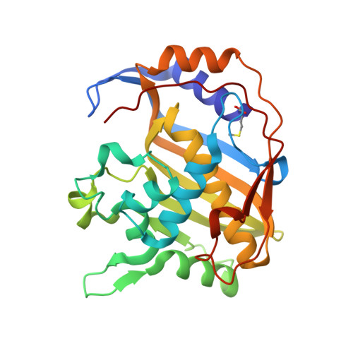
6NNR - PubMed Abstract:
Thymidylate synthase (TSase) produces the sole intracellular de novo source of thymidine (i.e., the DNA base T) and thus is a common target for antibiotic and anticancer drugs. Mg(2+) has been reported to affect TSase activity, but the mechanism of this interaction has not been investigated. Here we show that Mg(2+) binds to the surface of Escherichia coli TSase and affects the kinetics of hydride transfer at the interior active site (16 Å away). Examination of the crystal structures identifies a Mg(2+) near the glutamyl moiety of the folate cofactor, providing the first structural evidence for Mg(2+) binding to TSase. The kinetics and NMR relaxation experiments suggest that the weak binding of Mg(2+) to the protein surface stabilizes the closed conformation of the ternary enzyme complex and reduces the entropy of activation on the hydride transfer step. Mg(2+) accelerates the hydride transfer by ~7-fold but does not affect the magnitude or temperature dependence of the intrinsic kinetic isotope effect. These results suggest that Mg(2+) facilitates the protein motions that bring the hydride donor and acceptor together, but it does not change the tunneling ready state of the hydride transfer. These findings highlight how variations in cellular Mg(2+) concentration can modulate enzyme activity through long-range interactions in the protein, rather than binding at the active site. The interaction of Mg(2+) with the glutamyl tail of the folate cofactor and nonconserved residues of bacterial TSase may assist in designing antifolates with polyglutamyl substitutes as species-specific antibiotic drugs.
- Department of Chemistry, University of Iowa, Iowa City, Iowa 52242, USA.
Organizational Affiliation: