


'Catch and Release": a Variation of the Archetypal Nucleotidyl Transfer Reaction
Selvaraj, B., Kocaman, S., Trifas, M., Serpersu, E.H., Cuneo, M.J.(2020) ACS Catal
Experimental Data Snapshot
(2020) ACS Catal
Entity ID: 1 | |||||
|---|---|---|---|---|---|
| Molecule | Chains | Sequence Length | Organism | Details | Image |
| Kanamycin nucleotidyltransferase | 256 | Geobacillus stearothermophilus | Mutation(s): 0 EC: 2.7.7 | 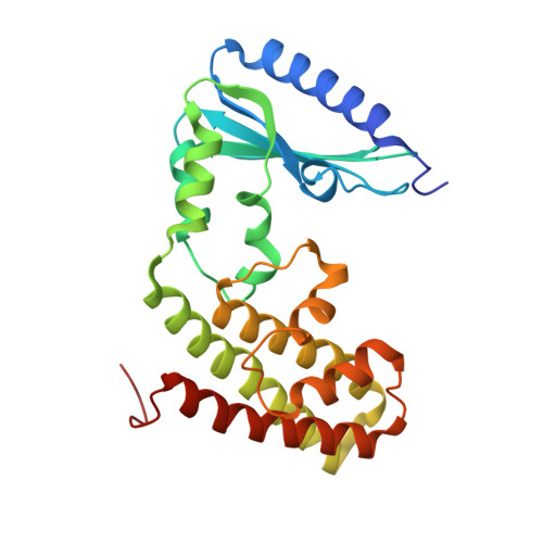 | |
UniProt | |||||
Entity Groups | |||||
| Sequence Clusters | 30% Identity50% Identity70% Identity90% Identity95% Identity100% Identity | ||||
| UniProt Group | P05057 | ||||
Sequence AnnotationsExpand | |||||
Reference Sequence | |||||
| Ligands 4 Unique | |||||
|---|---|---|---|---|---|
| ID | Chains | Name / Formula / InChI Key | 2D Diagram | 3D Interactions | |
| NMY Download:Ideal Coordinates CCD File | E [auth A] J [auth A] L [auth B] Q [auth C] V [auth C] | NEOMYCIN C23 H46 N6 O13 PGBHMTALBVVCIT-VCIWKGPPSA-N |  | ||
| AMP Download:Ideal Coordinates CCD File | F [auth A] K [auth A] M [auth B] R [auth C] W [auth C] | ADENOSINE MONOPHOSPHATE C10 H14 N5 O7 P UDMBCSSLTHHNCD-KQYNXXCUSA-N |  | ||
| PPV Download:Ideal Coordinates CCD File | G [auth A], N [auth B], S [auth C], Z [auth D] | PYROPHOSPHATE H4 O7 P2 XPPKVPWEQAFLFU-UHFFFAOYSA-N |  | ||
| MG Download:Ideal Coordinates CCD File | AA [auth D] BA [auth D] H [auth A] I [auth A] O [auth B] | MAGNESIUM ION Mg JLVVSXFLKOJNIY-UHFFFAOYSA-N |  | ||
| Length ( Å ) | Angle ( ˚ ) |
|---|---|
| a = 85.51 | α = 90 |
| b = 59.25 | β = 94.8 |
| c = 102.04 | γ = 90 |
| Software Name | Purpose |
|---|---|
| PHENIX | refinement |
| MOSFLM | data reduction |
| Aimless | data scaling |
| PHASER | phasing |
| Funding Organization | Location | Grant Number |
|---|---|---|
| National Science Foundation (NSF, United States) | United States | -- |
| Department of Energy (DOE, United States) | United States | -- |


