6NL9 | pdb_00006nl9

Crystal structure of de novo designed metal-controlled dimer of mutant B1 immunoglobulin-binding domain of Streptococcal Protein G (L12H, T16L, V29H, Y33H, N37L)-apo


Experimental Data Snapshot

  • Method: X-RAY DIFFRACTION
  • Resolution: 1.70 Å
  • R-Value Free: 
    0.224 (Depositor), 0.248 (DCC) 
  • R-Value Work: 
    0.186 (Depositor) 
  • R-Value Observed: 
    0.188 (Depositor) 

Starting Model: experimental
View more details

wwPDB Validation 3D Report Full Report

Validation slider image for 6NL9

This is version 1.4 of the entry. See complete history

Literature

Design of High-Affinity Metal-Controlled Protein Dimers.

Maniaci, B.Lipper, C.H.Anipindi, D.L.Erlandsen, H.Cole, J.L.Stec, B.Huxford, T.Love, J.J.

(2019) Biochemistry 58: 2199-2207

  • DOI: https://doi.org/10.1021/acs.biochem.9b00055
  • Primary Citation Related Structures: 
    6NL6, 6NL7, 6NL8, 6NL9, 6NLA, 6NLB

  • PubMed Abstract: 

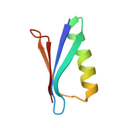
    The ability to precisely control protein complex formation has high utility in the expanding field of biomaterials. Driving protein-protein binding through metal-ligand bridging interactions is a promising method of achieving this goal. Furthermore, the capacity to precisely regulate both complex formation and dissociation enables additional control not available with constitutive protein complexes. Here we describe the design of three metal-controlled protein dimers that are completely monomeric in the absence of metal yet form high-affinity symmetric homodimers in the presence of zinc sulfate. The scaffold used for the designed dimers is the β1 domain of streptococcal protein G. In addition to forming high-affinity dimers in the presence of metal, the complexes also dissociate upon addition of EDTA. Biophysical characterization revealed that the proteins maintain relatively high thermal stability, bind with high affinity, and are completely monodisperse in the monomeric and dimeric states. High-resolution crystal structures revealed that the dimers adopt the target structure and that the designed metal-binding histidine residues successfully bind zinc and function to drive dimer formation.


  • Organizational Affiliation
    • Department of Chemistry and Biochemistry , San Diego State University , San Diego , California 92182 , United States.

Macromolecule Content 

  • Total Structure Weight: 25.08 kDa 
  • Atom Count: 1,938 
  • Modeled Residue Count: 224 
  • Deposited Residue Count: 224 
  • Unique protein chains: 1

Macromolecules

Find similar proteins by:|  3D Structure
Entity ID: 1
MoleculeChains  Sequence LengthOrganismDetailsImage
Immunoglobulin G-binding protein G
A, B, C, D
56StreptococcusMutation(s): 5 
Gene Names: spg
UniProt
Find proteins for P19909 (Streptococcus sp. group G)
Explore P19909 
Go to UniProtKB:  P19909
Entity Groups
Sequence Clusters30% Identity50% Identity70% Identity90% Identity95% Identity100% Identity
UniProt GroupP19909
Sequence Annotations
Expand
Reference Sequence

Experimental Data & Validation

Experimental Data

  • Method: X-RAY DIFFRACTION
  • Resolution: 1.70 Å
  • R-Value Free:  0.224 (Depositor), 0.248 (DCC) 
  • R-Value Work:  0.186 (Depositor) 
  • R-Value Observed: 0.188 (Depositor) 
Space Group: P 1 21 1
Unit Cell:
Length ( Å )Angle ( ˚ )
a = 45.817α = 90
b = 52.027β = 114.48
c = 50.148γ = 90
Software Package:
Software NamePurpose
REFMACrefinement
HKL-2000data reduction
HKL-2000data scaling
PHASERphasing

Structure Validation

View Full Validation Report


Entry History 

Deposition Data

Revision History  (Full details and data files)

  • Version 1.0: 2019-01-23
    Type: Initial release
  • Version 1.1: 2019-05-08
    Changes: Data collection, Database references
  • Version 1.2: 2019-05-15
    Changes: Data collection, Structure summary
  • Version 1.3: 2023-05-03
    Changes: Database references, Derived calculations, Structure summary
  • Version 1.4: 2023-10-25
    Changes: Data collection, Refinement description