


Crystal structure of Histidine--tRNA ligase from Elizabethkingia sp. CCUG 26117
Abendroth, J., Dranow, D.M., Lorimer, D.D., Horanyi, P.S., Edwards, T.E.To be published.
Experimental Data Snapshot
Starting Models: experimental
View more details
wwPDB Validation 3D Report Full Report

Entity ID: 1 | |||||
|---|---|---|---|---|---|
| Molecule | Chains | Sequence Length | Organism | Details | Image |
| Histidine--tRNA ligase | 457 | Elizabethkingia meningoseptica | Mutation(s): 0 Gene Names: hisS, NCTC10588_00401 EC: 6.1.1.21 | 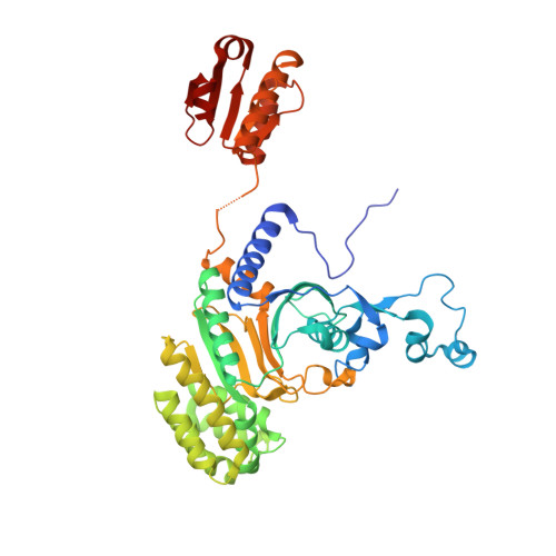 | |
| Length ( Å ) | Angle ( ˚ ) |
|---|---|
| a = 95.8 | α = 90 |
| b = 68.56 | β = 118.66 |
| c = 97.22 | γ = 90 |
| Software Name | Purpose |
|---|---|
| XDS | data reduction |
| XSCALE | data scaling |
| PHENIX | refinement |
| PDB_EXTRACT | data extraction |
| MoRDa | phasing |
| PHASER | phasing |
| PARROT | phasing |
| Coot | model building |


