6NEC

STRUCTURE OF RET PROTEIN TYROSINE KINASE DOMAIN IN COMPLEX WITH NINTEDANIB


Experimental Data Snapshot

  • Method: X-RAY DIFFRACTION
  • Resolution: 1.87 Å
  • R-Value Free: 0.232 
  • R-Value Work: 0.194 
  • R-Value Observed: 0.196 

wwPDB Validation   3D Report Full Report

Currently 6NEC does not have a validation slider image.


This is version 1.4 of the entry. See complete history


Literature

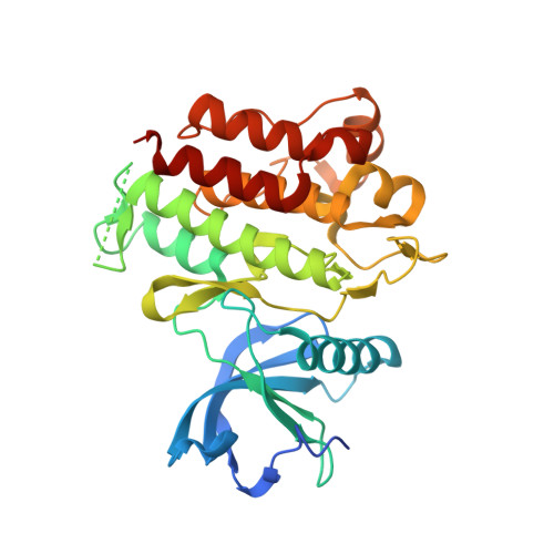
Structural basis of resistance of mutant RET protein-tyrosine kinase to its inhibitors nintedanib and vandetanib.

Terzyan, S.S.Shen, T.Liu, X.Huang, Q.Teng, P.Zhou, M.Hilberg, F.Cai, J.Mooers, B.H.M.Wu, J.

(2019) J Biol Chem 294: 10428-10437

  • DOI: https://doi.org/10.1074/jbc.RA119.007682
  • Primary Citation of Related Structures:  
    6NE7, 6NEC, 6NJA

  • PubMed Abstract: 

    RET is a transmembrane growth factor receptor. Aberrantly activated RET is found in several types of human cancer and is a target for treating RET aberration-associated cancer. Multiple clinically relevant RET protein-tyrosine kinase inhibitors (TKIs) have been identified, but how TKIs bind to RET is unknown except for vandetanib. Nintedanib is a RET TKI that inhibits the vandetanib-resistant RET(G810A) mutant. Here, we determined the X-ray co-crystal structure of RET kinase domain-nintedanib complex to 1.87 Å resolution and a RET(G810A) kinase domain crystal structure to 1.99 Å resolution. We also identified a vandetanib-resistant RET(L881V) mutation previously found in familial medullary thyroid carcinoma. Drug-sensitivity profiling of RET(L881V) revealed that it remains sensitive to nintedanib. The RET-nintedanib co-crystal structure disclosed that Leu-730 in RET engages in hydrophobic interactions with the piperazine, anilino, and phenyl groups of nintedanib, providing a structural basis for explaining that the p.L730V mutation identified in nine independently isolated cell lines resistant to nintedanib. Comparisons of RET-nintedanib, RET(G810A), and RET-vandetanib crystal structures suggested that the solvent-front Ala-810 makes hydrophobic contacts with a methyl group and aniline in nintedanib and blocks water access to two oxygen atoms of vandetanib, resulting in an energetic penalty for burying polar groups. Of note, even though the p.L881V mutation did not affect sensitivity to nintedanib, RET(L881V) was resistant to nintedanib analogs lacking a phenyl group. These results provide structural insights into resistance of RET mutants against the TKIs nintedanib and vandetanib.


  • Organizational Affiliation

    From the Departments of Biochemistry and Molecular Biology and.


Macromolecules
Find similar proteins by:  (by identity cutoff)  |  3D Structure
Entity ID: 1
MoleculeChains Sequence LengthOrganismDetailsImage
Proto-oncogene tyrosine-protein kinase receptor RetA,
B [auth C]
314Homo sapiensMutation(s): 0 
Gene Names: RETCDHF12CDHR16PTCRET51
EC: 2.7.10.1
UniProt & NIH Common Fund Data Resources
Find proteins for P07949 (Homo sapiens)
Explore P07949 
Go to UniProtKB:  P07949
PHAROS:  P07949
GTEx:  ENSG00000165731 
Entity Groups  
Sequence Clusters30% Identity50% Identity70% Identity90% Identity95% Identity100% Identity
UniProt GroupP07949
Sequence Annotations
Expand
  • Reference Sequence
Small Molecules
Ligands 2 Unique
IDChains Name / Formula / InChI Key2D Diagram3D Interactions
XIN
Query on XIN

Download Ideal Coordinates CCD File 
C [auth A],
F [auth C]
methyl (3Z)-3-{[(4-{methyl[(4-methylpiperazin-1-yl)acetyl]amino}phenyl)amino](phenyl)methylidene}-2-oxo-2,3-dihydro-1H-indole-6-carboxylate
C31 H33 N5 O4
XZXHXSATPCNXJR-ZIADKAODSA-N
FMT
Query on FMT

Download Ideal Coordinates CCD File 
D [auth A],
E [auth C]
FORMIC ACID
C H2 O2
BDAGIHXWWSANSR-UHFFFAOYSA-N
Binding Affinity Annotations 
IDSourceBinding Affinity
XIN BindingDB:  6NEC Kd: min: 9.6, max: 31 (nM) from 4 assay(s)
Experimental Data & Validation

Experimental Data

  • Method: X-RAY DIFFRACTION
  • Resolution: 1.87 Å
  • R-Value Free: 0.232 
  • R-Value Work: 0.194 
  • R-Value Observed: 0.196 
  • Space Group: P 1 21 1
Unit Cell:
Length ( Å )Angle ( ˚ )
a = 50.587α = 90
b = 80.295β = 100.52
c = 79.926γ = 90
Software Package:
Software NamePurpose
REFMACrefinement
HKL-3000data reduction
HKL-3000data scaling
MOLREPphasing

Structure Validation

View Full Validation Report

Currently 6NEC does not have a validation slider image.


Entry History & Funding Information

Deposition Data


Funding OrganizationLocationGrant Number
National Institutes of Health/National Human Genome Research Institute (NIH/NHGRI)United StatesP20GM103640

Revision History  (Full details and data files)

  • Version 1.0: 2019-06-05
    Type: Initial release
  • Version 1.1: 2019-07-17
    Changes: Data collection, Database references
  • Version 1.2: 2019-09-25
    Changes: Data collection, Experimental preparation
  • Version 1.3: 2019-12-18
    Changes: Author supporting evidence
  • Version 1.4: 2023-10-11
    Changes: Data collection, Database references, Refinement description