6NDY

Vps4 with Cyclic Peptide Bound in the Central Pore


Experimental Data Snapshot

  • Method: ELECTRON MICROSCOPY
  • Resolution: 3.60 Å
  • Aggregation State: PARTICLE 
  • Reconstruction Method: SINGLE PARTICLE 

wwPDB Validation   3D Report Full Report


This is version 1.2 of the entry. See complete history


Literature

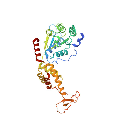
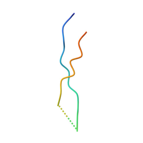
Structure of Vps4 with circular peptides and implications for translocation of two polypeptide chains by AAA+ ATPases.

Han, H.Fulcher, J.M.Dandey, V.P.Iwasa, J.H.Sundquist, W.I.Kay, M.S.Shen, P.S.Hill, C.P.

(2019) Elife 8

  • DOI: https://doi.org/10.7554/eLife.44071
  • Primary Citation of Related Structures:  
    6NDY, 6OO2

  • PubMed Abstract: 

    Many AAA+ ATPases form hexamers that unfold protein substrates by translocating them through their central pore. Multiple structures have shown how a helical assembly of subunits binds a single strand of substrate, and indicate that translocation results from the ATP-driven movement of subunits from one end of the helical assembly to the other end. To understand how more complex substrates are bound and translocated, we demonstrated that linear and cyclic versions of peptides bind to the S. cerevisiae AAA+ ATPase Vps4 with similar affinities, and determined cryo-EM structures of cyclic peptide complexes. The peptides bind in a hairpin conformation, with one primary strand equivalent to the single chain peptide ligands, while the second strand returns through the translocation pore without making intimate contacts with Vps4. These observations indicate a general mechanism by which AAA+ ATPases may translocate a variety of substrates that include extended chains, hairpins, and crosslinked polypeptide chains.


  • Organizational Affiliation

    Department of Biochemistry, University of Utah, Salt Lake City, United States.


Macromolecules
Find similar proteins by:  (by identity cutoff)  |  3D Structure
Entity ID: 1
MoleculeChains Sequence LengthOrganismDetailsImage
Vacuolar protein sorting-associated protein 4
A, B, C, D, E
337Saccharomyces cerevisiaeMutation(s): 0 
Gene Names: VPS4CSC1DID6END13GRD13VPL4VPT10YPR173CP9705.10
UniProt
Find proteins for P52917 (Saccharomyces cerevisiae (strain ATCC 204508 / S288c))
Explore P52917 
Go to UniProtKB:  P52917
Entity Groups  
Sequence Clusters30% Identity50% Identity70% Identity90% Identity95% Identity100% Identity
UniProt GroupP52917
Sequence Annotations
Expand
  • Reference Sequence
Find similar proteins by:  (by identity cutoff)  |  3D Structure
Entity ID: 2
MoleculeChains Sequence LengthOrganismDetailsImage
Designed Cyclic PeptideF [auth G]30synthetic constructMutation(s): 0 
Entity Groups  
Sequence Clusters30% Identity50% Identity70% Identity90% Identity95% Identity100% Identity
Sequence Annotations
Expand
  • Reference Sequence
Small Molecules
Ligands 3 Unique
IDChains Name / Formula / InChI Key2D Diagram3D Interactions
ADP
Query on ADP

Download Ideal Coordinates CCD File 
G [auth A],
J [auth B],
M [auth C],
P [auth D],
R [auth E]
ADENOSINE-5'-DIPHOSPHATE
C10 H15 N5 O10 P2
XTWYTFMLZFPYCI-KQYNXXCUSA-N
BEF
Query on BEF

Download Ideal Coordinates CCD File 
H [auth A],
K [auth B],
N [auth C]
BERYLLIUM TRIFLUORIDE ION
Be F3
OGIAHMCCNXDTIE-UHFFFAOYSA-K
MG
Query on MG

Download Ideal Coordinates CCD File 
I [auth A],
L [auth B],
O [auth C],
Q [auth D]
MAGNESIUM ION
Mg
JLVVSXFLKOJNIY-UHFFFAOYSA-N
Experimental Data & Validation

Experimental Data

  • Method: ELECTRON MICROSCOPY
  • Resolution: 3.60 Å
  • Aggregation State: PARTICLE 
  • Reconstruction Method: SINGLE PARTICLE 
EM Software:
TaskSoftware PackageVersion
RECONSTRUCTIONRELION

Structure Validation

View Full Validation Report


Entry History & Funding Information

Deposition Data


Funding OrganizationLocationGrant Number
National Institutes of Health/National Human Genome Research Institute (NIH/NHGRI)United StatesP50 GM082545
National Institutes of Health/National Human Genome Research Institute (NIH/NHGRI)United StatesR01 GM112080
National Institutes of Health/National Human Genome Research Institute (NIH/NHGRI)United StatesP41 GM103310

Revision History  (Full details and data files)

  • Version 1.0: 2019-08-21
    Type: Initial release
  • Version 1.1: 2019-12-18
    Changes: Author supporting evidence, Other
  • Version 1.2: 2024-03-20
    Changes: Data collection, Database references, Derived calculations