


Crystal structure of a LPMO from Kitasatospora papulosa
Correa, T.L.C., Tomazini Jr., A., Murakami, M.T.To be published.
Experimental Data Snapshot
Starting Model: experimental
View more details
wwPDB Validation 3D Report Full Report

Entity ID: 1 | |||||
|---|---|---|---|---|---|
| Molecule | Chains | Sequence Length | Organism | Details | Image |
| lytic polysaccharide monooxygenase | 192 | Kitasatospora papulosa | Mutation(s): 0 | 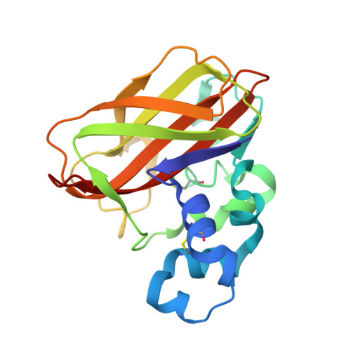 | |
| Length ( Å ) | Angle ( ˚ ) |
|---|---|
| a = 37.207 | α = 90 |
| b = 109.574 | β = 113.46 |
| c = 42.775 | γ = 90 |
| Software Name | Purpose |
|---|---|
| PHENIX | refinement |
| XDS | data reduction |
| XSCALE | data scaling |
| PHASER | phasing |
| Funding Organization | Location | Grant Number |
|---|---|---|
| Sao Paulo Research Foundation (FAPESP) | Brazil | 15/26982-0 |


