6NAF

De novo designed homo-trimeric amantadine-binding protein


Experimental Data Snapshot

  • Method: X-RAY DIFFRACTION
  • Resolution: 1.92 Å
  • R-Value Free: 0.204 
  • R-Value Work: 0.175 
  • R-Value Observed: 0.178 

  • Method: NEUTRON DIFFRACTION
  • Resolution: 2.50 Å
  • R-Value Free: 0.318 
  • R-Value Work: 0.277 
  • R-Value Observed: 0.280 

wwPDB Validation   3D Report Full Report


This is version 1.2 of the entry. See complete history


Literature

De novo design of a homo-trimeric amantadine-binding protein.

Park, J.Selvaraj, B.McShan, A.C.Boyken, S.E.Wei, K.Y.Oberdorfer, G.DeGrado, W.Sgourakis, N.G.Cuneo, M.J.Myles, D.A.Baker, D.

(2019) Elife 8

  • DOI: https://doi.org/10.7554/eLife.47839
  • Primary Citation of Related Structures:  
    6N9H, 6NAF

  • PubMed Abstract: 

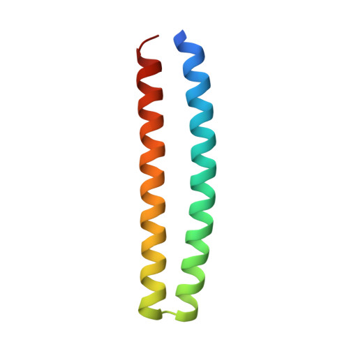
    The computational design of a symmetric protein homo-oligomer that binds a symmetry-matched small molecule larger than a metal ion has not yet been achieved. We used de novo protein design to create a homo-trimeric protein that binds the C 3 symmetric small molecule drug amantadine with each protein monomer making identical interactions with each face of the small molecule. Solution NMR data show that the protein has regular three-fold symmetry and undergoes localized structural changes upon ligand binding. A high-resolution X-ray structure reveals a close overall match to the design model with the exception of water molecules in the amantadine binding site not included in the Rosetta design calculations, and a neutron structure provides experimental validation of the computationally designed hydrogen-bond networks. Exploration of approaches to generate a small molecule inducible homo-trimerization system based on the design highlight challenges that must be overcome to computationally design such systems.


  • Organizational Affiliation

    Department of Biochemistry, University of Washington, Seattle, United States.


Macromolecules
Find similar proteins by:  (by identity cutoff)  |  3D Structure
Entity ID: 1
MoleculeChains Sequence LengthOrganismDetailsImage
amantadine-binding protein80synthetic constructMutation(s): 0 
Entity Groups  
Sequence Clusters30% Identity50% Identity70% Identity90% Identity95% Identity100% Identity
Sequence Annotations
Expand
  • Reference Sequence
Small Molecules
Ligands 2 Unique
IDChains Name / Formula / InChI Key2D Diagram3D Interactions
308 (Subject of Investigation/LOI)
Query on 308

Download Ideal Coordinates CCD File 
B [auth A](3S,5S,7S)-tricyclo[3.3.1.1~3,7~]decan-1-amine
C10 H17 N
DKNWSYNQZKUICI-CHIWXEEVSA-N
NA
Query on NA

Download Ideal Coordinates CCD File 
C [auth A]SODIUM ION
Na
FKNQFGJONOIPTF-UHFFFAOYSA-N
Experimental Data & Validation

Experimental Data

  • Method: X-RAY DIFFRACTION
  • Resolution: 1.92 Å
  • R-Value Free: 0.204 
  • R-Value Work: 0.175 
  • R-Value Observed: 0.178 
  • Space Group: P 63
  • Method: NEUTRON DIFFRACTION
  • Resolution: 2.50 Å
  • R-Value Free: 0.318 
  • R-Value Work: 0.277 
  • R-Value Observed: 0.280 
  • Space Group: P 63
Unit Cell:
Length ( Å )Angle ( ˚ )
a = 50.61α = 90
b = 50.61β = 90
c = 68.82γ = 120
Software Package:
Software NamePurpose
PHENIXrefinement
LAUEGENdata reduction
LSCALEdata scaling
PHASERphasing

Structure Validation

View Full Validation Report


Entry History & Funding Information

Deposition Data


Funding OrganizationLocationGrant Number
Department of Energy (DOE, United States)United States--

Revision History  (Full details and data files)

  • Version 1.0: 2019-12-18
    Type: Initial release
  • Version 1.1: 2020-01-01
    Changes: Database references
  • Version 1.2: 2023-10-25
    Changes: Data collection, Database references, Derived calculations, Refinement description