6NAE | pdb_00006nae

Crystal Structure of Ebola zaire GP protein with bound ARN0074898


Experimental Data Snapshot

  • Method: X-RAY DIFFRACTION
  • Resolution: 2.75 Å
  • R-Value Free: 
    0.240 (Depositor), 0.242 (DCC) 
  • R-Value Work: 
    0.193 (Depositor), 0.196 (DCC) 
  • R-Value Observed: 
    0.197 (Depositor) 

Starting Model: experimental
View more details

wwPDB Validation 3D Report Full Report

Validation slider image for 6NAE

Ligand Structure Quality Assessment 


This is version 2.2 of the entry. See complete history

Literature

Discovery of Adamantane Carboxamides as Ebola Virus Cell Entry and Glycoprotein Inhibitors.

Plewe, M.B.Sokolova, N.V.Gantla, V.R.Brown, E.R.Naik, S.Fetsko, A.Lorimer, D.D.Dranow, D.M.Smutney, H.Bullen, J.Sidhu, R.Master, A.Wang, J.Kallel, E.A.Zhang, L.Kalveram, B.Freiberg, A.N.Henkel, G.McCormack, K.

(2020) ACS Med Chem Lett 11: 1160-1167

  • DOI: https://doi.org/10.1021/acsmedchemlett.0c00025
  • Primary Citation Related Structures: 
    6NAE

  • PubMed Abstract: 

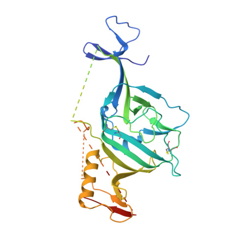
    We identified and explored the structure-activity-relationship (SAR) of an adamantane carboxamide chemical series of Ebola virus (EBOV) inhibitors. Selected analogs exhibited half-maximal inhibitory concentrations (EC 50 values) of ∼10-15 nM in vesicular stomatitis virus (VSV) pseudotyped EBOV (pEBOV) infectivity assays, low hundred nanomolar EC 50 activity against wild type EBOV, aqueous solubility >20 mg/mL, and attractive metabolic stability in human and nonhuman liver microsomes. X-ray cocrystallographic characterizations of a lead compound with the EBOV glycoprotein (GP) established the EBOV GP as a target for direct compound inhibitory activity and further provided relevant structural models that may assist in identifying optimized therapeutic candidates.


  • Organizational Affiliation
    • Arisan Therapeutics, 11189 Sorrento Valley Road, Suite 104, San Diego, California 92121, United States.

Macromolecule Content 

  • Total Structure Weight: 57.85 kDa 
  • Atom Count: 2,972 
  • Modeled Residue Count: 355 
  • Deposited Residue Count: 498 
  • Unique protein chains: 2

Macromolecules

Find similar proteins by:|  3D Structure
Entity ID: 1
MoleculeChains  Sequence LengthOrganismDetailsImage
Envelope glycoprotein,Envelope glycoprotein,Envelope glycoprotein330Ebola virus - Mayinga, Zaire, 1976Mutation(s): 1 
Gene Names: GP
UniProt
Find proteins for Q05320 (Zaire ebolavirus (strain Mayinga-76))
Explore Q05320 
Go to UniProtKB:  Q05320
Entity Groups
Sequence Clusters30% Identity50% Identity70% Identity90% Identity95% Identity100% Identity
UniProt GroupQ05320
Glycosylation
Glycosylation Sites: 4
Sequence Annotations
Expand
Reference Sequence
Find similar proteins by:|  3D Structure
Entity ID: 2
MoleculeChains  Sequence LengthOrganismDetailsImage
Envelope glycoprotein168Ebola virus - Mayinga, Zaire, 1976Mutation(s): 1 
Gene Names: GP
UniProt
Find proteins for Q05320 (Zaire ebolavirus (strain Mayinga-76))
Explore Q05320 
Go to UniProtKB:  Q05320
Entity Groups
Sequence Clusters30% Identity50% Identity70% Identity90% Identity95% Identity100% Identity
UniProt GroupQ05320
Glycosylation
Glycosylation Sites: 1
Sequence Annotations
Expand
Reference Sequence

Oligosaccharides

Help  
Entity ID: 3
MoleculeChains Length2D Diagram GlycosylationD Interactions
alpha-D-mannopyranose-(1-3)-[alpha-D-mannopyranose-(1-6)]beta-D-mannopyranose-(1-4)-2-acetamido-2-deoxy-beta-D-glucopyranose-(1-4)-2-acetamido-2-deoxy-beta-D-glucopyranose
C
5N-Glycosylation
Glycosylation Resources
GlyTouCan: G22768VO
GlyCosmos: G22768VO
GlyGen: G22768VO

Small Molecules

Ligands 3 Unique
IDChains Name / Formula / InChI Key2D Diagram3D Interactions
KHG

Query on KHG


Download:Ideal Coordinates CCD File
K [auth A](1S,3R,5R,7S)-N-(trans-4-aminocyclohexyl)-3-methyl-5-phenyltricyclo[3.3.1.1~3,7~]decane-1-carboxamide
C24 H34 N2 O
TZNRCHUCVIMHIH-UCWUHLBISA-N
NAG

Query on NAG


Download:Ideal Coordinates CCD File
D [auth A],
E [auth A],
F [auth A],
G [auth A]
2-acetamido-2-deoxy-beta-D-glucopyranose
C8 H15 N O6
OVRNDRQMDRJTHS-FMDGEEDCSA-N
GOL

Query on GOL


Download:Ideal Coordinates CCD File
H [auth A],
I [auth A],
J [auth A],
L [auth A],
M [auth B]
GLYCEROL
C3 H8 O3
PEDCQBHIVMGVHV-UHFFFAOYSA-N

Experimental Data & Validation

Experimental Data

  • Method: X-RAY DIFFRACTION
  • Resolution: 2.75 Å
  • R-Value Free:  0.240 (Depositor), 0.242 (DCC) 
  • R-Value Work:  0.193 (Depositor), 0.196 (DCC) 
  • R-Value Observed: 0.197 (Depositor) 
Space Group: H 3 2
Unit Cell:
Length ( Å )Angle ( ˚ )
a = 113.25α = 90
b = 113.25β = 90
c = 307.21γ = 120
Software Package:
Software NamePurpose
PHENIXrefinement
XDSdata reduction
XSCALEdata scaling
PHASERphasing
PDB_EXTRACTdata extraction

Structure Validation

View Full Validation Report


Ligand Structure Quality Assessment 


Entry History 

Deposition Data

Revision History  (Full details and data files)

  • Version 1.0: 2019-12-11
    Type: Initial release
  • Version 1.1: 2019-12-25
    Changes: Database references
  • Version 1.2: 2020-07-01
    Changes: Data collection, Database references
  • Version 2.0: 2020-07-29
    Type: Remediation
    Reason: Carbohydrate remediation
    Changes: Atomic model, Data collection, Derived calculations, Structure summary
  • Version 2.1: 2023-10-11
    Changes: Data collection, Database references, Derived calculations, Refinement description, Structure summary
  • Version 2.2: 2024-11-20
    Changes: Structure summary