6N6R

Crystal structure of ABIN-1 UBAN in complex with two M1-linked di-ubiquitins


Experimental Data Snapshot

  • Method: X-RAY DIFFRACTION
  • Resolution: 1.95 Å
  • R-Value Free: 0.278 
  • R-Value Work: 0.243 
  • R-Value Observed: 0.245 

wwPDB Validation   3D Report Full Report


This is version 1.2 of the entry. See complete history


Literature

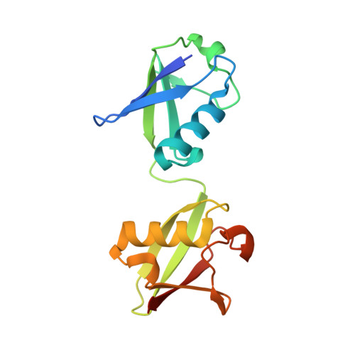
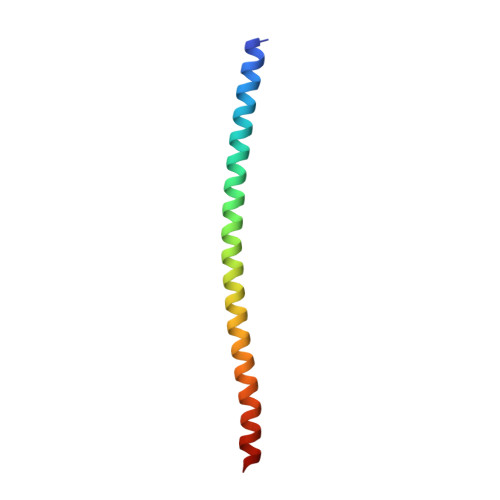
Molecular Recognition of M1-Linked Ubiquitin Chains by Native and Phosphorylated UBAN Domains.

Herhaus, L.van den Bedem, H.Tang, S.Maslennikov, I.Wakatsuki, S.Dikic, I.Rahighi, S.

(2019) J Mol Biol 431: 3146-3156

  • DOI: https://doi.org/10.1016/j.jmb.2019.06.012
  • Primary Citation of Related Structures:  
    6N5M, 6N6R, 6N6S

  • PubMed Abstract: 

    Although the Ub-binding domain in ABIN proteins and NEMO (UBAN) is highly conserved, UBAN-containing proteins exhibit different Ub-binding properties, resulting in their diverse biological roles. Post-translational modifications further control UBAN domain specificity for poly-Ub chains. However, precisely, how the UBAN domain structurally confers such functional diversity remains poorly understood. Here we report crystal structures of ABIN-1 alone and in complex with one or two M1-linked di-Ub chains. ABIN-1 UBAN forms a homo-dimer that provides two symmetrical Ub-binding sites on either side of the coiled-coil structure. Moreover, crystal structures of ABIN1 UBAN in complex with di-Ub chains reveal a concentration-dependency of UBAN/di-Ub binding stoichiometry. Analysis of UBAN/M1-linked di-Ub binding characteristics indicates that phosphorylated S473 in OPTN and its corresponding phospho-mimetic residue in ABIN-1 (E484) are essential for high affinity interactions with M1-linked Ub chains. Also, a phospho-mimetic mutation of A303 in NEMO, corresponding to S473 of OPTN, increases binding affinity for M1-linked Ub chains. These findings are in line with the diverse physiological roles of UBAN domains, as phosphorylation of OPTN UBAN is required to enhance its binding to Ub during mitophagy.


  • Organizational Affiliation

    Institute of Biochemistry II, Goethe University School of Medicine, Theodor-Stern-Kai 7, 60590 Frankfurt am Main, Germany.


Macromolecules
Find similar proteins by:  (by identity cutoff)  |  3D Structure
Entity ID: 1
MoleculeChains Sequence LengthOrganismDetailsImage
UbiquitinA,
D [auth C]
157Homo sapiensMutation(s): 0 
Gene Names: UBC
UniProt & NIH Common Fund Data Resources
Find proteins for P0CG48 (Homo sapiens)
Explore P0CG48 
Go to UniProtKB:  P0CG48
PHAROS:  P0CG48
GTEx:  ENSG00000150991 
Entity Groups  
Sequence Clusters30% Identity50% Identity70% Identity90% Identity95% Identity100% Identity
UniProt GroupP0CG48
Sequence Annotations
Expand
  • Reference Sequence
Find similar proteins by:  (by identity cutoff)  |  3D Structure
Entity ID: 2
MoleculeChains Sequence LengthOrganismDetailsImage
TNFAIP3-interacting protein 1B,
C [auth D]
70Mus musculusMutation(s): 0 
Gene Names: Tnip1AbinNaf1
UniProt & NIH Common Fund Data Resources
Find proteins for Q9WUU8 (Mus musculus)
Explore Q9WUU8 
Go to UniProtKB:  Q9WUU8
IMPC:  MGI:1926194
Entity Groups  
Sequence Clusters30% Identity50% Identity70% Identity90% Identity95% Identity100% Identity
UniProt GroupQ9WUU8
Sequence Annotations
Expand
  • Reference Sequence
Experimental Data & Validation

Experimental Data

  • Method: X-RAY DIFFRACTION
  • Resolution: 1.95 Å
  • R-Value Free: 0.278 
  • R-Value Work: 0.243 
  • R-Value Observed: 0.245 
  • Space Group: P 21 21 21
Unit Cell:
Length ( Å )Angle ( ˚ )
a = 52.9α = 90
b = 88.18β = 90
c = 105.9γ = 90
Software Package:
Software NamePurpose
REFMACrefinement
MOSFLMdata reduction
Aimlessdata scaling
MOLREPphasing

Structure Validation

View Full Validation Report


Entry History 

Deposition Data

Revision History  (Full details and data files)

  • Version 1.0: 2019-07-17
    Type: Initial release
  • Version 1.1: 2019-08-28
    Changes: Data collection, Database references
  • Version 1.2: 2024-03-13
    Changes: Data collection, Database references, Refinement description