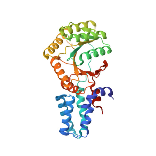
6N6N

FtsY-NG high-resolution


Experimental Data Snapshot

  • Method: X-RAY DIFFRACTION
  • Resolution: 1.88 Å
  • R-Value Free: 0.243 
  • R-Value Work: 0.195 
  • R-Value Observed: 0.197 

Starting Model: experimental
View more details

wwPDB Validation   3D Report Full Report


Ligand Structure Quality Assessment 


This is version 1.3 of the entry. See complete history


Literature

Structural insights into the G-loop dynamics of E. coli FtsY NG domain.

Faoro, C.Ataide, S.F.

(2019) J Struct Biol 208: 107387-107387

  • DOI: https://doi.org/10.1016/j.jsb.2019.09.004
  • Primary Citation of Related Structures:  
    6N5I, 6N5J, 6N6N, 6N9B, 6NC1, 6NC4

  • PubMed Abstract: 

    The bacterial signal recognition particle (SRP) receptor, FtsY, participates with the SRP in co-translation targeting of proteins. Multiple crystal structures of the NG domain of E. coli FtsY NG have been determined at high-resolution (1.22-1.88 Å), in the nucleotide-free (apo) form as well as bound to GDP and non-hydrolysable GTP analogues. The combination of high-resolution and multiple solved structures of FtsY NG in different states revealed a new sensor-relay system of this unique GTPase receptor. A nucleotide sensing function of the P-loop assists FtsY NG in nucleotide-binding and contributes to modulate nucleotide binding properties in SRP GTPases. A reorganization of the other G-loops and the insertion binding domain (IBD) is observed only upon transition from a diphosphate to a triphosphate nucleotide. The role of a magnesium ion during the GDP and GTP-bound states has also been observed. The binding of magnesium in the nucleotide site causes the reorientation of the β- and γ- phosphate groups toward the jaws of the P-loop and stabilizes the binding of the nucleotide, creating a network of hydrogen and water-bridge interactions.


  • Organizational Affiliation

    School of Life and Environmental Sciences, The University of Sydney, Sydney 2006, Australia.


Macromolecules
Find similar proteins by:  (by identity cutoff)  |  3D Structure
Entity ID: 1
MoleculeChains Sequence LengthOrganismDetailsImage
Signal recognition particle receptor FtsY
A, B
303Escherichia coli K-12Mutation(s): 0 
Gene Names: ftsYb3464JW3429
UniProt
Find proteins for P10121 (Escherichia coli (strain K12))
Explore P10121 
Go to UniProtKB:  P10121
Entity Groups  
Sequence Clusters30% Identity50% Identity70% Identity90% Identity95% Identity100% Identity
UniProt GroupP10121
Sequence Annotations
Expand
  • Reference Sequence
Small Molecules
Ligands 4 Unique
IDChains Name / Formula / InChI Key2D Diagram3D Interactions
GCP (Subject of Investigation/LOI)
Query on GCP

Download Ideal Coordinates CCD File 
C [auth A],
H [auth B]
PHOSPHOMETHYLPHOSPHONIC ACID GUANYLATE ESTER
C11 H18 N5 O13 P3
PHBDHXOBFUBCJD-KQYNXXCUSA-N
GOL
Query on GOL

Download Ideal Coordinates CCD File 
I [auth B]GLYCEROL
C3 H8 O3
PEDCQBHIVMGVHV-UHFFFAOYSA-N
ACT
Query on ACT

Download Ideal Coordinates CCD File 
G [auth A]ACETATE ION
C2 H3 O2
QTBSBXVTEAMEQO-UHFFFAOYSA-M
MG (Subject of Investigation/LOI)
Query on MG

Download Ideal Coordinates CCD File 
D [auth A],
E [auth A],
F [auth A],
J [auth B],
K [auth B]
MAGNESIUM ION
Mg
JLVVSXFLKOJNIY-UHFFFAOYSA-N
Experimental Data & Validation

Experimental Data

  • Method: X-RAY DIFFRACTION
  • Resolution: 1.88 Å
  • R-Value Free: 0.243 
  • R-Value Work: 0.195 
  • R-Value Observed: 0.197 
  • Space Group: P 1 21 1
Unit Cell:
Length ( Å )Angle ( ˚ )
a = 35.42α = 90
b = 76.883β = 92.54
c = 106.935γ = 90
Software Package:
Software NamePurpose
PHENIXrefinement
XDSdata reduction
Aimlessdata scaling
MOLREPphasing

Structure Validation

View Full Validation Report


Ligand Structure Quality Assessment 


Entry History 

Deposition Data

Revision History  (Full details and data files)

  • Version 1.0: 2019-10-09
    Type: Initial release
  • Version 1.1: 2019-12-04
    Changes: Database references
  • Version 1.2: 2020-03-18
    Changes: Database references
  • Version 1.3: 2023-10-11
    Changes: Data collection, Database references, Derived calculations, Refinement description