


Crystal structure of histone lysine methyltransferase SmyD2 in complex with polyethylene glycol
Perry, E., Yang, Z.To be published.
Experimental Data Snapshot
Starting Model: experimental
View more details
Entity ID: 1 | |||||
|---|---|---|---|---|---|
| Molecule | Chains | Sequence Length | Organism | Details | Image |
| N-lysine methyltransferase SMYD2 | 433 | Homo sapiens | Mutation(s): 0 Gene Names: SMYD2, KMT3C EC: 2.1.1 (PDB Primary Data), 2.1.1.43 (PDB Primary Data), 2.1.1.354 (UniProt) | 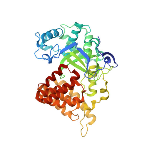 | |
UniProt & NIH Common Fund Data Resources | |||||
PHAROS: Q9NRG4 GTEx: ENSG00000143499 | |||||
Entity Groups | |||||
| Sequence Clusters | 30% Identity50% Identity70% Identity90% Identity95% Identity100% Identity | ||||
| UniProt Group | Q9NRG4 | ||||
Sequence AnnotationsExpand | |||||
Reference Sequence | |||||
| Ligands 5 Unique | |||||
|---|---|---|---|---|---|
| ID | Chains | Name / Formula / InChI Key | 2D Diagram | 3D Interactions | |
| 12P Download:Ideal Coordinates CCD File | C [auth A] | DODECAETHYLENE GLYCOL C24 H50 O13 WRZXKWFJEFFURH-UHFFFAOYSA-N |  | ||
| SAH Download:Ideal Coordinates CCD File | B [auth A] | S-ADENOSYL-L-HOMOCYSTEINE C14 H20 N6 O5 S ZJUKTBDSGOFHSH-WFMPWKQPSA-N |  | ||
| ZN Download:Ideal Coordinates CCD File | K [auth A], L [auth A], M [auth A] | ZINC ION Zn PTFCDOFLOPIGGS-UHFFFAOYSA-N |  | ||
| NI Download:Ideal Coordinates CCD File | D [auth A], E [auth A] | NICKEL (II) ION Ni VEQPNABPJHWNSG-UHFFFAOYSA-N |  | ||
| EOH Download:Ideal Coordinates CCD File | F [auth A], G [auth A], H [auth A], I [auth A], J [auth A] | ETHANOL C2 H6 O LFQSCWFLJHTTHZ-UHFFFAOYSA-N |  | ||
| Length ( Å ) | Angle ( ˚ ) |
|---|---|
| a = 151.84 | α = 90 |
| b = 151.84 | β = 90 |
| c = 53.74 | γ = 90 |
| Software Name | Purpose |
|---|---|
| PHENIX | refinement |
| XDS | data reduction |
| Aimless | data scaling |
| PHENIX | phasing |
| autoPROC | data collection |


