6N17

Crystal structure of Tdp1 catalytic domain in complex with compound XZ577


Experimental Data Snapshot

  • Method: X-RAY DIFFRACTION
  • Resolution: 1.64 Å
  • R-Value Free: 0.211 
  • R-Value Work: 0.184 
  • R-Value Observed: 0.184 

wwPDB Validation   3D Report Full Report

Currently 6N17 does not have a validation slider image.


This is version 1.2 of the entry. See complete history


Literature

Identification of a ligand binding hot spot and structural motifs replicating aspects of tyrosyl-DNA phosphodiesterase I (TDP1) phosphoryl recognition by crystallographic fragment cocktail screening.

Lountos, G.T.Zhao, X.Z.Kiselev, E.Tropea, J.E.Needle, D.Pommier, Y.Burke, T.R.Waugh, D.S.

(2019) Nucleic Acids Res 47: 10134-10150

  • DOI: https://doi.org/10.1093/nar/gkz515
  • Primary Citation of Related Structures:  
    6DHU, 6DIE, 6DIH, 6DIM, 6DJD, 6DJE, 6DJF, 6DJG, 6DJH, 6DJI, 6DJJ, 6MJ5, 6N17, 6N19

  • PubMed Abstract: 

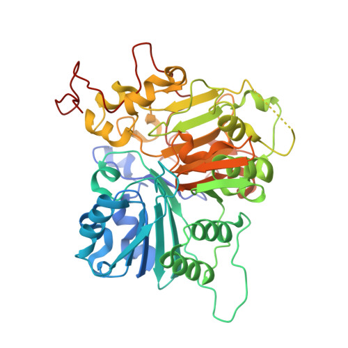
    Tyrosyl DNA-phosphodiesterase I (TDP1) repairs type IB topoisomerase (TOP1) cleavage complexes generated by TOP1 inhibitors commonly used as anticancer agents. TDP1 also removes DNA 3' end blocking lesions generated by chain-terminating nucleosides and alkylating agents, and base oxidation both in the nuclear and mitochondrial genomes. Combination therapy with TDP1 inhibitors is proposed to synergize with topoisomerase targeting drugs to enhance selectivity against cancer cells exhibiting deficiencies in parallel DNA repair pathways. A crystallographic fragment screening campaign against the catalytic domain of TDP1 was conducted to identify new lead compounds. Crystal structures revealed two fragments that bind to the TDP1 active site and exhibit inhibitory activity against TDP1. These fragments occupy a similar position in the TDP1 active site as seen in prior crystal structures of TDP1 with bound vanadate, a transition state mimic. Using structural insights into fragment binding, several fragment derivatives have been prepared and evaluated in biochemical assays. These results demonstrate that fragment-based methods can be a highly feasible approach toward the discovery of small-molecule chemical scaffolds to target TDP1, and for the first time, we provide co-crystal structures of small molecule inhibitors bound to TDP1, which could serve for the rational development of medicinal TDP1 inhibitors.


  • Organizational Affiliation

    Basic Science Program, Frederick National Laboratory for Cancer Research, Frederick, MD 21702, USA.


Macromolecules
Find similar proteins by:  (by identity cutoff)  |  3D Structure
Entity ID: 1
MoleculeChains Sequence LengthOrganismDetailsImage
Tyrosyl-DNA phosphodiesterase 1
A, B
461Homo sapiensMutation(s): 0 
Gene Names: TDP1
EC: 3.1.4
UniProt & NIH Common Fund Data Resources
Find proteins for Q9NUW8 (Homo sapiens)
Explore Q9NUW8 
Go to UniProtKB:  Q9NUW8
PHAROS:  Q9NUW8
GTEx:  ENSG00000042088 
Entity Groups  
Sequence Clusters30% Identity50% Identity70% Identity90% Identity95% Identity100% Identity
UniProt GroupQ9NUW8
Sequence Annotations
Expand
  • Reference Sequence
Small Molecules
Ligands 3 Unique
IDChains Name / Formula / InChI Key2D Diagram3D Interactions
K8Y
Query on K8Y

Download Ideal Coordinates CCD File 
C [auth A],
G [auth B]
4-[(3-carboxypropanoyl)amino]benzene-1,2-dicarboxylic acid
C12 H11 N O7
JVZSGTXFIVTSIN-UHFFFAOYSA-N
DMS
Query on DMS

Download Ideal Coordinates CCD File 
F [auth A]DIMETHYL SULFOXIDE
C2 H6 O S
IAZDPXIOMUYVGZ-UHFFFAOYSA-N
EDO
Query on EDO

Download Ideal Coordinates CCD File 
D [auth A],
E [auth A],
H [auth B],
I [auth B],
J [auth B]
1,2-ETHANEDIOL
C2 H6 O2
LYCAIKOWRPUZTN-UHFFFAOYSA-N
Binding Affinity Annotations 
IDSourceBinding Affinity
K8Y Binding MOAD:  6N17 IC50: 1.01e+6 (nM) from 1 assay(s)
Experimental Data & Validation

Experimental Data

  • Method: X-RAY DIFFRACTION
  • Resolution: 1.64 Å
  • R-Value Free: 0.211 
  • R-Value Work: 0.184 
  • R-Value Observed: 0.184 
  • Space Group: P 21 21 21
Unit Cell:
Length ( Å )Angle ( ˚ )
a = 50.049α = 90
b = 105.411β = 90
c = 194.093γ = 90
Software Package:
Software NamePurpose
PHENIXrefinement
HKL-3000data reduction
HKL-3000data scaling
PHASERphasing

Structure Validation

View Full Validation Report

Currently 6N17 does not have a validation slider image.


Entry History 

Deposition Data

Revision History  (Full details and data files)

  • Version 1.0: 2019-07-03
    Type: Initial release
  • Version 1.1: 2019-11-13
    Changes: Data collection, Database references
  • Version 1.2: 2023-10-11
    Changes: Data collection, Database references, Refinement description