6N14 | pdb_00006n14

Phosphoserine BlaC, Class A serine beta-lactamase from Mycobacterium tuberculosis


Experimental Data Snapshot

  • Method: X-RAY DIFFRACTION
  • Resolution: 1.52 Å
  • R-Value Free: 
    0.169 (Depositor), 0.170 (DCC) 
  • R-Value Work: 
    0.155 (Depositor), 0.155 (DCC) 
  • R-Value Observed: 
    0.155 (Depositor) 

Starting Model: experimental
View more details

wwPDB Validation 3D Report Full Report

Validation slider image for 6N14

This is version 1.2 of the entry. See complete history

Literature

Crystal Structure of Phosphoserine BlaC fromMycobacterium tuberculosisInactivated by Bis(Benzoyl) Phosphate.

Moural, T.W.White, D.S.Choy, C.J.Kang, C.Berkman, C.E.

(2019) Int J Mol Sci 20

  • DOI: https://doi.org/10.3390/ijms20133247
  • Primary Citation Related Structures: 
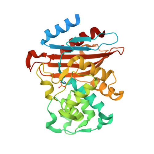
    6N14

  • PubMed Abstract: 

    Mycobacterium tuberculosis , the pathogen responsible for tuberculosis (TB), is the leading cause of death from infectious disease worldwide. The class A serine β-lactamase BlaC confers Mycobacterium tuberculosis resistance to conventional β-lactam antibiotics. As the primary mechanism of bacterial resistance to β-lactam antibiotics, the expression of a β-lactamase by Mycobacterium tuberculosis results in hydrolysis of the β-lactam ring and deactivation of these antibiotics. In this study, we conducted protein X-ray crystallographic analysis of the inactivation of BlaC, upon exposure to the inhibitor bis(benzoyl) phosphate. Crystal structure data confirms that serine β-lactamase is phosphorylated at the catalytic serine residue (Ser-70) by this phosphate-based inactivator. This new crystallographic evidence suggests a mechanism for phosphorylation of BlaC inhibition by bis(benzoyl) phosphate over acylation. Additionally, we confirmed that bis(benzoyl) phosphate inactivated BlaC in a time-dependent manner.


  • Organizational Affiliation
    • Department of Chemistry, Washington State University, Pullman, WA 99164, USA.

Macromolecule Content 

  • Total Structure Weight: 32.89 kDa 
  • Atom Count: 2,328 
  • Modeled Residue Count: 265 
  • Deposited Residue Count: 307 
  • Unique protein chains: 1

Macromolecules

Find similar proteins by:|  3D Structure
Entity ID: 1
MoleculeChains  Sequence LengthOrganismDetailsImage
Beta-lactamase307Mycobacterium tuberculosisMutation(s): 0 
Gene Names: blaCERS027646_02769
EC: 3.5.2.6
UniProt
Find proteins for P9WKD3 (Mycobacterium tuberculosis (strain ATCC 25618 / H37Rv))
Explore P9WKD3 
Go to UniProtKB:  P9WKD3
Entity Groups
Sequence Clusters30% Identity50% Identity70% Identity90% Identity95% Identity100% Identity
UniProt GroupP9WKD3
Sequence Annotations
Expand
Reference Sequence

Small Molecules

Modified Residues  1 Unique
IDChains TypeFormula2D DiagramParent
SEP
Query on SEP
A
L-PEPTIDE LINKINGC3 H8 N O6 PSER

Experimental Data & Validation

Experimental Data

  • Method: X-RAY DIFFRACTION
  • Resolution: 1.52 Å
  • R-Value Free:  0.169 (Depositor), 0.170 (DCC) 
  • R-Value Work:  0.155 (Depositor), 0.155 (DCC) 
  • R-Value Observed: 0.155 (Depositor) 
Space Group: P 21 21 21
Unit Cell:
Length ( Å )Angle ( ˚ )
a = 49.84α = 90
b = 68.037β = 90
c = 75.454γ = 90
Software Package:
Software NamePurpose
HKL-2000data scaling
PHENIXrefinement
PDB_EXTRACTdata extraction
HKL-2000data reduction
PHENIXphasing

Structure Validation

View Full Validation Report


Entry History 

Deposition Data

Revision History  (Full details and data files)

  • Version 1.0: 2019-08-28
    Type: Initial release
  • Version 1.1: 2023-10-11
    Changes: Data collection, Database references, Refinement description
  • Version 1.2: 2024-11-13
    Changes: Structure summary