6MYU | pdb_00006myu

Avian mitochondrial complex II crystallized in the presence of HQNO


Experimental Data Snapshot

  • Method: X-RAY DIFFRACTION
  • Resolution: 1.97 Å
  • R-Value Free: 
    0.223 (Depositor), 0.224 (DCC) 
  • R-Value Work: 
    0.199 (Depositor), 0.199 (DCC) 
  • R-Value Observed: 
    0.199 (Depositor) 

Starting Model: experimental
View more details

wwPDB Validation 3D Report Full Report

Validation slider image for 6MYU

Ligand Structure Quality Assessment 


This is version 1.3 of the entry. See complete history

Literature

Crystallographic investigation of the ubiquinone binding site of respiratory Complex II and its inhibitors.

Huang, L.S.Lummen, P.Berry, E.A.

(2021) Biochim Biophys Acta Proteins Proteom 1869: 140679-140679

  • DOI: https://doi.org/10.1016/j.bbapap.2021.140679
  • Primary Citation Related Structures: 
    6MYO, 6MYP, 6MYQ, 6MYR, 6MYS, 6MYT, 6MYU

  • PubMed Abstract: 

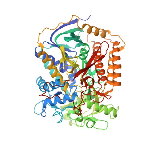
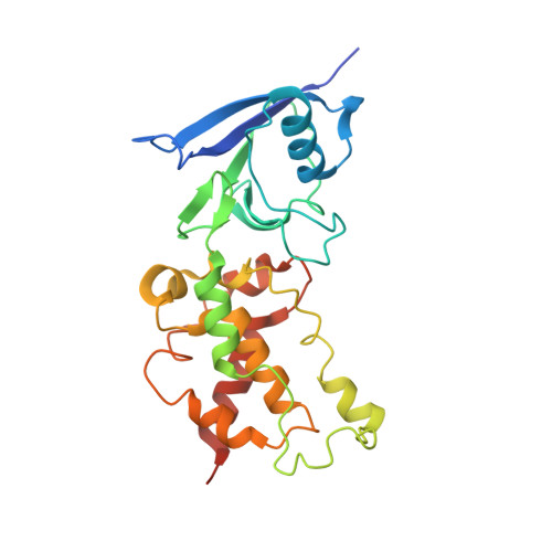
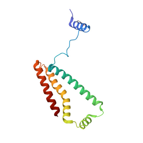
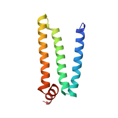
    The quinone binding site (Q-site) of Mitochondrial Complex II (succinate-ubiquinone oxidoreductase) is the target for a number of inhibitors useful for elucidating the mechanism of the enzyme. Some of these have been developed as fungicides or pesticides, and species-specific Q-site inhibitors may be useful against human pathogens. We report structures of chicken Complex II with six different Q-site inhibitors bound, at resolutions 2.0-2.4 Å. These structures show the common interactions between the inhibitors and their binding site. In every case a carbonyl or hydroxyl oxygen of the inhibitor is H-bonded to Tyr58 in subunit SdhD and Trp173 in subunit SdhB. Two of the inhibitors H-bond Ser39 in subunit SdhC directly, while two others do so via a water molecule. There is a distinct cavity that accepts the 2-substituent of the carboxylate ring in flutolanil and related inhibitors. A hydrophobic "tail pocket" opens to receive a side-chain of intermediate-length inhibitors. Shorter inhibitors fit entirely within the main binding cleft, while the long hydrophobic side chains of ferulenol and atpenin A5 protrude out of the cleft into the bulk lipid region, as presumably does that of ubiquinone. Comparison of mitochondrial and Escherichia coli Complex II shows a rotation of the membrane-anchor subunits by 7° relative to the iron‑sulfur protein. This rotation alters the geometry of the Q-site and the H-bonding pattern of SdhB:His216 and SdhD:Asp57. This conformational difference, rather than any active-site mutation, may be responsible for the different inhibitor sensitivity of the bacterial enzyme.


  • Organizational Affiliation
    • Biochemistry and Molecular Biology, SUNY Upstate Medical University, 750 E. Adams Street, Syracuse, N.Y 13210, USA.

Macromolecule Content 

  • Total Structure Weight: 127.09 kDa 
  • Atom Count: 9,421 
  • Modeled Residue Count: 1,092 
  • Deposited Residue Count: 1,116 
  • Unique protein chains: 4

Macromolecules

Find similar proteins by:|  3D Structure
Entity ID: 1
MoleculeChains  Sequence LengthOrganismDetailsImage
Succinate dehydrogenase [ubiquinone] flavoprotein subunit, mitochondrial621Gallus gallusMutation(s): 0 
EC: 1.3.5.1 (PDB Primary Data), 1.1.5 (UniProt)
Membrane Entity: Yes 
UniProt
Find proteins for Q9YHT1 (Gallus gallus)
Explore Q9YHT1 
Go to UniProtKB:  Q9YHT1
Entity Groups
Sequence Clusters30% Identity50% Identity70% Identity90% Identity95% Identity100% Identity
UniProt GroupQ9YHT1
Sequence Annotations
Expand
Reference Sequence
Find similar proteins by:|  3D Structure
Entity ID: 2
MoleculeChains  Sequence LengthOrganismDetailsImage
Succinate dehydrogenase [ubiquinone] iron-sulfur subunit, mitochondrial252Gallus gallusMutation(s): 0 
EC: 1.3.5.1 (PDB Primary Data), 1.1.5 (UniProt)
Membrane Entity: Yes 
UniProt
Find proteins for Q9YHT2 (Gallus gallus)
Explore Q9YHT2 
Go to UniProtKB:  Q9YHT2
Entity Groups
Sequence Clusters30% Identity50% Identity70% Identity90% Identity95% Identity100% Identity
UniProt GroupQ9YHT2
Sequence Annotations
Expand
Reference Sequence
Find similar proteins by:|  3D Structure
Entity ID: 3
MoleculeChains  Sequence LengthOrganismDetailsImage
Succinate dehydrogenase cytochrome b, large subunit140Gallus gallusMutation(s): 0 
EC: 1.3.5.1
Membrane Entity: Yes 
UniProt
Find proteins for D0VWW3 (Gallus gallus)
Explore D0VWW3 
Go to UniProtKB:  D0VWW3
Entity Groups
Sequence Clusters30% Identity50% Identity70% Identity90% Identity95% Identity100% Identity
UniProt GroupD0VWW3
Sequence Annotations
Expand
Reference Sequence
Find similar proteins by:|  3D Structure
Entity ID: 4
MoleculeChains  Sequence LengthOrganismDetailsImage
Succinate dehydrogenase [ubiquinone] cytochrome b small subunit, mitochondrial103Gallus gallusMutation(s): 0 
Membrane Entity: Yes 
UniProt
Find proteins for Q5ZIS0 (Gallus gallus)
Explore Q5ZIS0 
Go to UniProtKB:  Q5ZIS0
Entity Groups
Sequence Clusters30% Identity50% Identity70% Identity90% Identity95% Identity100% Identity
UniProt GroupQ5ZIS0
Sequence Annotations
Expand
Reference Sequence

Small Molecules

Ligands 11 Unique
IDChains Name / Formula / InChI Key2D Diagram3D Interactions
FAD

Query on FAD


Download:Ideal Coordinates CCD File
E [auth A]FLAVIN-ADENINE DINUCLEOTIDE
C27 H33 N9 O15 P2
VWWQXMAJTJZDQX-UYBVJOGSSA-N
3PE

Query on 3PE


Download:Ideal Coordinates CCD File
BC [auth D]1,2-Distearoyl-sn-glycerophosphoethanolamine
C41 H82 N O8 P
LVNGJLRDBYCPGB-LDLOPFEMSA-N
HEM

Query on HEM


Download:Ideal Coordinates CCD File
FB [auth C]PROTOPORPHYRIN IX CONTAINING FE
C34 H32 Fe N4 O4
KABFMIBPWCXCRK-RGGAHWMASA-L
UMQ

Query on UMQ


Download:Ideal Coordinates CCD File
GB [auth C]UNDECYL-MALTOSIDE
C23 H44 O11
UYEMNFYVTFDKRG-ZNGNCRBCSA-N
SF4

Query on SF4


Download:Ideal Coordinates CCD File
MA [auth B]IRON/SULFUR CLUSTER
Fe4 S4
LJBDFODJNLIPKO-UHFFFAOYSA-N
F3S

Query on F3S


Download:Ideal Coordinates CCD File
NA [auth B]FE3-S4 CLUSTER
Fe3 S4
FCXHZBQOKRZXKS-UHFFFAOYSA-N
FES

Query on FES


Download:Ideal Coordinates CCD File
LA [auth B]FE2/S2 (INORGANIC) CLUSTER
Fe2 S2
NIXDOXVAJZFRNF-UHFFFAOYSA-N
OAA

Query on OAA


Download:Ideal Coordinates CCD File
F [auth A]OXALOACETATE ION
C4 H3 O5
KHPXUQMNIQBQEV-UHFFFAOYSA-M
PEG

Query on PEG


Download:Ideal Coordinates CCD File
I [auth A]DI(HYDROXYETHYL)ETHER
C4 H10 O3
MTHSVFCYNBDYFN-UHFFFAOYSA-N
K

Query on K


Download:Ideal Coordinates CCD File
G [auth A],
OA [auth B]
POTASSIUM ION
K
NPYPAHLBTDXSSS-UHFFFAOYSA-N
UNL

Query on UNL


Download:Ideal Coordinates CCD File
AA [auth A]
AB [auth B]
AC [auth C]
BA [auth A]
BB [auth B]
AA [auth A],
AB [auth B],
AC [auth C],
BA [auth A],
BB [auth B],
CA [auth A],
CB [auth B],
CC [auth D],
DA [auth A],
DB [auth B],
DC [auth D],
EA [auth A],
EB [auth C],
EC [auth D],
FA [auth A],
FC [auth D],
GA [auth A],
GC [auth D],
H [auth A],
HA [auth A],
HB [auth C],
HC [auth D],
IA [auth A],
IB [auth C],
IC [auth D],
J [auth A],
JA [auth A],
JB [auth C],
JC [auth D],
K [auth A],
KA [auth A],
KB [auth C],
KC [auth D],
L [auth A],
LB [auth C],
LC [auth D],
M [auth A],
MB [auth C],
MC [auth D],
N [auth A],
NB [auth C],
NC [auth D],
O [auth A],
OB [auth C],
OC [auth D],
P [auth A],
PA [auth B],
PB [auth C],
PC [auth D],
Q [auth A],
QA [auth B],
QB [auth C],
QC [auth D],
R [auth A],
RA [auth B],
RB [auth C],
S [auth A],
SA [auth B],
SB [auth C],
T [auth A],
TA [auth B],
TB [auth C],
U [auth A],
UA [auth B],
UB [auth C],
V [auth A],
VA [auth B],
VB [auth C],
W [auth A],
WA [auth B],
WB [auth C],
X [auth A],
XA [auth B],
XB [auth C],
Y [auth A],
YA [auth B],
YB [auth C],
Z [auth A],
ZA [auth B],
ZB [auth C]
Unknown ligand
NPYPAHLBTDXSSS-UHFFFAOYSA-N
Modified Residues  1 Unique
IDChains TypeFormula2D DiagramParent
MLZ
Query on MLZ
C
L-PEPTIDE LINKINGC7 H16 N2 O2LYS

Experimental Data & Validation

Experimental Data

  • Method: X-RAY DIFFRACTION
  • Resolution: 1.97 Å
  • R-Value Free:  0.223 (Depositor), 0.224 (DCC) 
  • R-Value Work:  0.199 (Depositor), 0.199 (DCC) 
  • R-Value Observed: 0.199 (Depositor) 
Space Group: P 21 21 21
Unit Cell:
Length ( Å )Angle ( ˚ )
a = 70.029α = 90
b = 83.958β = 90
c = 290.54γ = 90
Software Package:
Software NamePurpose
PHENIXrefinement
DENZOdata reduction
SCALEPACKdata scaling
PHENIXphasing

Structure Validation

View Full Validation Report


Ligand Structure Quality Assessment 


Entry History 

& Funding Information

Deposition Data


Funding OrganizationLocationGrant Number
Department of Health & Human Services (HHS)United States1R01GM062563

Revision History  (Full details and data files)

  • Version 1.0: 2019-11-06
    Type: Initial release
  • Version 1.1: 2020-01-01
    Changes: Author supporting evidence
  • Version 1.2: 2021-07-07
    Changes: Database references, Derived calculations, Structure summary
  • Version 1.3: 2023-10-11
    Changes: Data collection, Database references, Refinement description