6MWR | pdb_00006mwr

Recognition of MHC-like molecule


Experimental Data Snapshot

  • Method: X-RAY DIFFRACTION
  • Resolution: 3.30 Å
  • R-Value Free: 
    0.261 (Depositor), 0.299 (DCC) 
  • R-Value Work: 
    0.238 (Depositor), 0.276 (DCC) 
  • R-Value Observed: 
    0.239 (Depositor) 

Starting Models: experimental
View more details

wwPDB Validation 3D Report Full Report

Validation slider image for 6MWR

Ligand Structure Quality Assessment 


This is version 1.4 of the entry. See complete history. 

Literature

A class of gamma delta T cell receptors recognize the underside of the antigen-presenting molecule MR1.

Le Nours, J., Gherardin, N.A., Ramarathinam, S.H., Awad, W., Wiede, F., Gully, B.S., Khandokar, Y., Praveena, T., Wubben, J.M., Sandow, J.J., Webb, A.I., von Borstel, A., Rice, M.T., Redmond, S.J., Seneviratna, R., Sandoval-Romero, M.L., Li, S., Souter, M.N.T., Eckle, S.B.G., Corbett, A.J., Reid, H.H., Liu, L., Fairlie, D.P., Giles, E.M., Westall, G.P., Tothill, R.W., Davey, M.S., Berry, R., Tiganis, T., McCluskey, J., Pellicci, D.G., Purcell, A.W., Uldrich, A.P., Godfrey, D.I., Rossjohn, J.

(2019) Science 366: 1522-1527

  • DOI: https://doi.org/10.1126/science.aav3900
  • Primary Citation Related Structures: 
    6MWR

  • PubMed Abstract: 

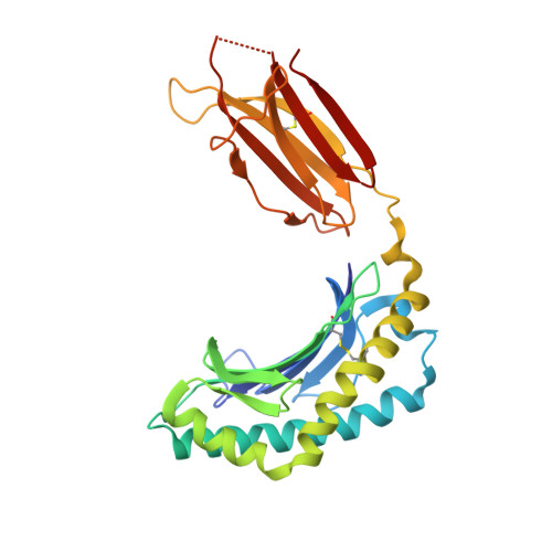
    T cell receptors (TCRs) recognize antigens presented by major histocompatibility complex (MHC) and MHC class I-like molecules. We describe a diverse population of human γδ T cells isolated from peripheral blood and tissues that exhibit autoreactivity to the monomorphic MHC-related protein 1 (MR1). The crystal structure of a γδTCR-MR1-antigen complex starkly contrasts with all other TCR-MHC and TCR-MHC-I-like complex structures. Namely, the γδTCR binds underneath the MR1 antigen-binding cleft, where contacts are dominated by the MR1 α3 domain. A similar pattern of reactivity was observed for diverse MR1-restricted γδTCRs from multiple individuals. Accordingly, we simultaneously report MR1 as a ligand for human γδ T cells and redefine the parameters for TCR recognition.


  • Organizational Affiliation: 
    • Infection and Immunity Program and Department of Biochemistry and Molecular Biology, Biomedicine Discovery Institute, Monash University, Clayton, Victoria 3800, Australia.

Macromolecule Content 

  • Total Structure Weight: 98.14 kDa 
  • Atom Count: 5,970 
  • Modeled Residue Count: 789 
  • Deposited Residue Count: 850 
  • Unique protein chains: 4

Macromolecules

Find similar proteins by:|  3D Structure
Entity ID: 1
MoleculeChains  Sequence LengthOrganismDetailsImage
Major histocompatibility complex class I-related gene protein271Homo sapiensMutation(s): 0 
Gene Names: MR1
UniProt & NIH Common Fund Data Resources
Find proteins for Q95460 (Homo sapiens)
Explore Q95460 
Go to UniProtKB:  Q95460
PHAROS:  Q95460
GTEx:  ENSG00000153029 
Entity Groups
Sequence Clusters30% Identity50% Identity70% Identity90% Identity95% Identity100% Identity
UniProt GroupQ95460
Sequence Annotations
Expand
Reference Sequence
Find similar proteins by:|  3D Structure
Entity ID: 2
MoleculeChains  Sequence LengthOrganismDetailsImage
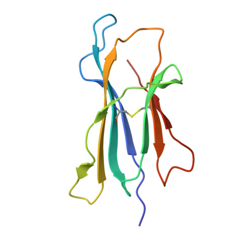
Beta-2-microglobulin99Homo sapiensMutation(s): 0 
Gene Names: B2M, CDABP0092, HDCMA22P
UniProt & NIH Common Fund Data Resources
Find proteins for P61769 (Homo sapiens)
Explore P61769 
Go to UniProtKB:  P61769
PHAROS:  P61769
GTEx:  ENSG00000166710 
Entity Groups
Sequence Clusters30% Identity50% Identity70% Identity90% Identity95% Identity100% Identity
UniProt GroupP61769
Sequence Annotations
Expand
Reference Sequence
Find similar proteins by:|  3D Structure
Entity ID: 3
MoleculeChains  Sequence LengthOrganismDetailsImage
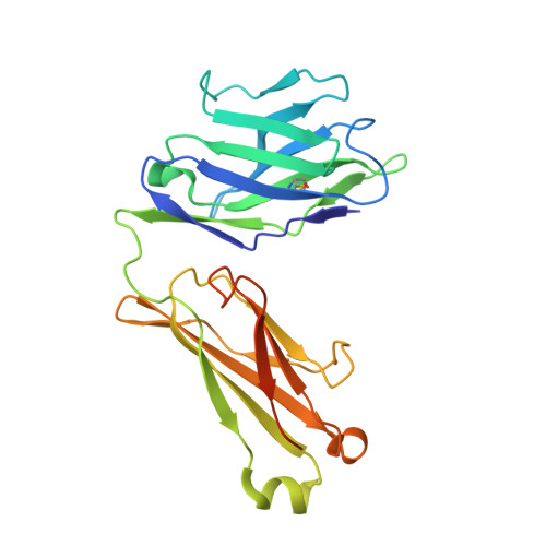
G7 Gamma chain T cell receptor244Homo sapiensMutation(s): 0 
Entity Groups
Sequence Clusters30% Identity50% Identity70% Identity90% Identity95% Identity100% Identity
Sequence Annotations
Expand
Reference Sequence
Find similar proteins by:|  3D Structure
Entity ID: 4
MoleculeChains  Sequence LengthOrganismDetailsImage
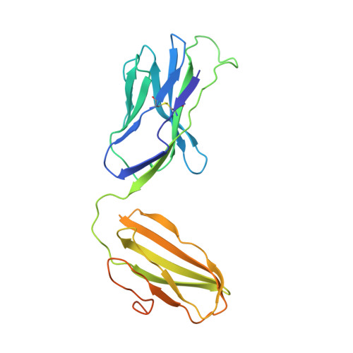
Delta chain of T cell receptor236Homo sapiensMutation(s): 0 
Entity Groups
Sequence Clusters30% Identity50% Identity70% Identity90% Identity95% Identity100% Identity
Glycosylation
Glycosylation Sites: 1
Sequence Annotations
Expand
Reference Sequence

Small Molecules

Ligands 2 Unique
IDChains Name / Formula / InChI Key2D Diagram3D Interactions
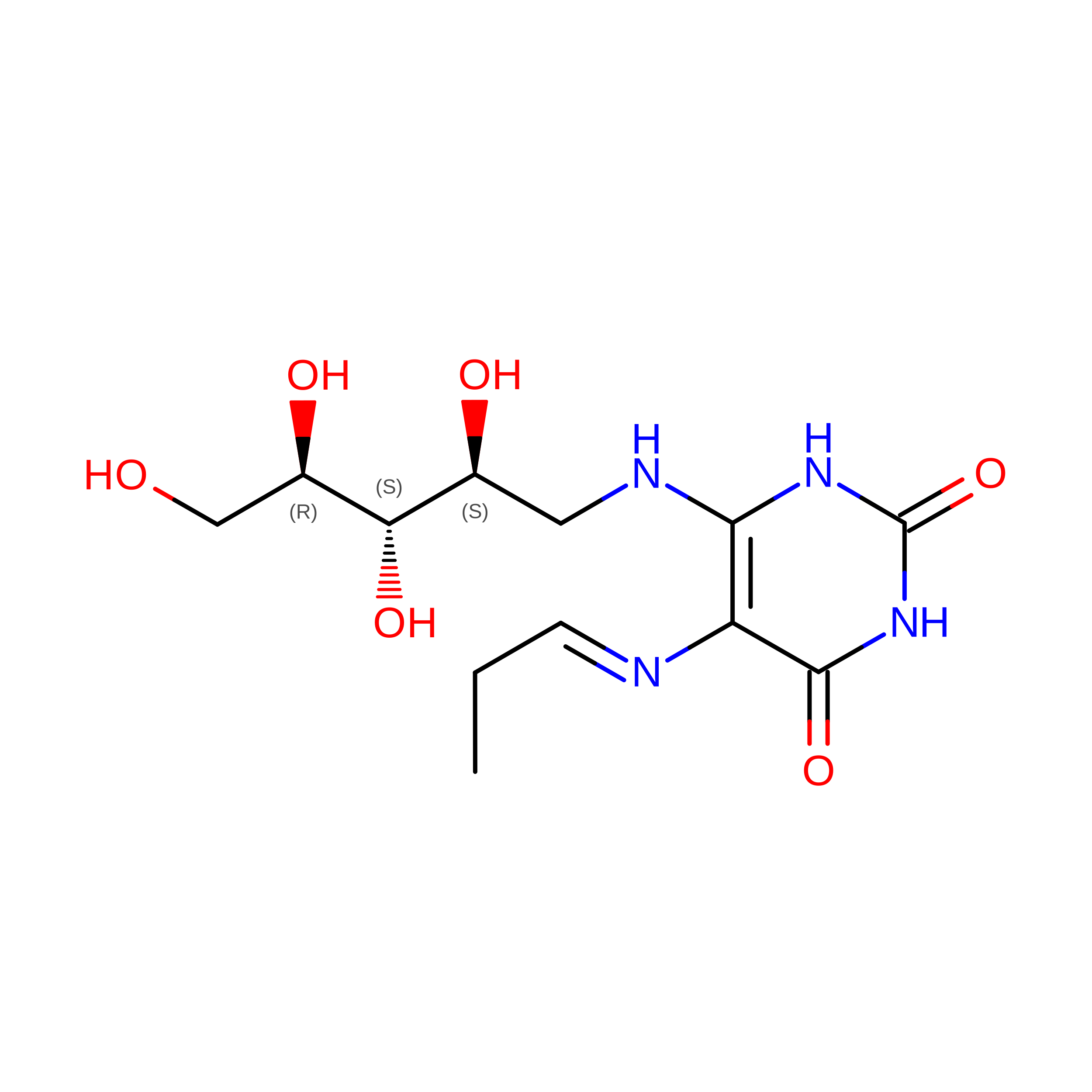
2LJ

Query on 2LJ


Download:Ideal Coordinates CCD File
E [auth A]1-deoxy-1-({2,6-dioxo-5-[(E)-propylideneamino]-1,2,3,6-tetrahydropyrimidin-4-yl}amino)-D-ribitol
C12 H20 N4 O6
YCMPUNANLDFPQG-FHZGFTDOSA-N
NAG

Query on NAG


Download:Ideal Coordinates CCD File
F [auth D]2-acetamido-2-deoxy-beta-D-glucopyranose
C8 H15 N O6
OVRNDRQMDRJTHS-FMDGEEDCSA-N

Experimental Data & Validation

Experimental Data

  • Method: X-RAY DIFFRACTION
  • Resolution: 3.30 Å
  • R-Value Free:  0.261 (Depositor), 0.299 (DCC) 
  • R-Value Work:  0.238 (Depositor), 0.276 (DCC) 
  • R-Value Observed: 0.239 (Depositor) 
Space Group: P 43 21 2
Unit Cell:
Length ( Å )Angle ( ˚ )
a = 88.39α = 90
b = 88.39β = 90
c = 257.15γ = 90
Software Package:
Software NamePurpose
BUSTERrefinement
XDSdata reduction
SCALAdata scaling
PHASERphasing

Structure Validation

View Full Validation Report


Ligand Structure Quality Assessment 


Entry History 

Deposition Data

Revision History  (Full details and data files)

  • Version 1.0: 2019-12-18
    Type: Initial release
  • Version 1.1: 2020-07-08
    Changes: Data collection, Database references
  • Version 1.2: 2020-07-29
    Type: Remediation
    Reason: Carbohydrate remediation
    Changes: Data collection, Derived calculations, Structure summary
  • Version 1.3: 2023-10-11
    Changes: Data collection, Database references, Refinement description, Structure summary
  • Version 1.4: 2024-10-23
    Changes: Structure summary