Discovery and Characterization of FMN-Binding beta-Glucuronidases in the Human Gut Microbiome.
Pellock, S.J., Walton, W.G., Ervin, S.M., Torres-Rivera, D., Creekmore, B.C., Bergan, G., Dunn, Z.D., Li, B., Tripathy, A., Redinbo, M.R.(2019) J Mol Biology 431: 970-980
- PubMed: 30658055 Search on PubMedSearch on PubMed Central
- DOI: https://doi.org/10.1016/j.jmb.2019.01.013
- Primary Citation Related Structures:
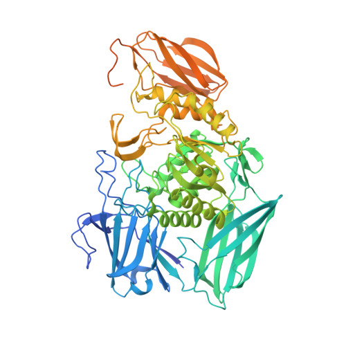
6MVF, 6MVG, 6MVH - PubMed Abstract:
The human gut microbiota encodes β-glucuronidases (GUSs) that play key roles in health and disease via the metabolism of glucuronate-containing carbohydrates and drugs. Hundreds of putative bacterial GUS enzymes have been identified by metagenomic analysis of the human gut microbiome, but less than 10% have characterized structures and functions. Here we describe a set of unique gut microbial GUS enzymes that bind flavin mononucleotide (FMN). First, we show using mass spectrometry, isothermal titration calorimetry, and x-ray crystallography that a purified GUS from the gut commensal microbe Faecalibacterium prausnitzii binds to FMN on a surface groove located 30 Å away from the active site. Second, utilizing structural and functional data from this FMN-binding GUS, we analyzed the 279 unique GUS sequences from the Human Microbiome Project database and identified 14 putative FMN-binding GUSs. We characterized four of these hits and solved the structure of two, the GUSs from Ruminococcus gnavus and Roseburia hominis, which confirmed that these are FMN binders. Third, binding and kinetic analysis of the FMN-binding site mutants of these five GUSs show that they utilize a conserved site to bind FMN that is not essential for GUS activity, but can affect K M . Lastly, a comprehensive structural review of the PDB reveals that the FMN-binding site employed by these enzymes is unlike any structurally characterized FMN binders to date. These findings reveal the first instance of an FMN-binding glycoside hydrolase and suggest a potential link between FMN and carbohydrate metabolism in the human gut microbiota.
- Department of Chemistry, University of North Carolina at Chapel Hill, Chapel Hill, NC 27599, USA.
Organizational Affiliation: