6MV7 | pdb_00006mv7

Crystal structure of RNAse 6


Experimental Data Snapshot

  • Method: X-RAY DIFFRACTION
  • Resolution: 2.59 Å
  • R-Value Free: 
    0.252 (Depositor), 0.259 (DCC) 
  • R-Value Work: 
    0.217 (Depositor), 0.217 (DCC) 
  • R-Value Observed: 
    0.220 (Depositor) 

Starting Model: experimental
View more details

wwPDB Validation 3D Report Full Report

Validation slider image for 6MV7

Ligand Structure Quality Assessment 


This is version 2.3 of the entry. See complete history

Literature

Insights into Structural and Dynamical Changes Experienced by Human RNase 6 upon Ligand Binding.

Narayanan, C.Bernard, D.N.Letourneau, M.Gagnon, J.Gagne, D.Bafna, K.Calmettes, C.Couture, J.F.Agarwal, P.K.Doucet, N.

(2020) Biochemistry 59: 755-765

  • DOI: https://doi.org/10.1021/acs.biochem.9b00888
  • Primary Citation Related Structures: 
    6MV6, 6MV7

  • PubMed Abstract: 

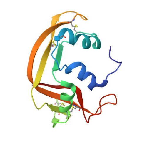
    Ribonuclease 6 (RNase 6) is one of eight catalytically active human pancreatic-type RNases that belong to a superfamily of rapidly evolving enzymes. Like some of its human homologues, RNase 6 exhibits host defense properties such as antiviral and antibacterial activities. Recently solved crystal structures of this enzyme in its nucleotide-free form show the conservation of the prototypical kidney-shaped fold preserved among vertebrate RNases, in addition to revealing the presence of a unique secondary active site. In this study, we determine the structural and conformational properties experienced by RNase 6 upon binding to substrate and product analogues. We present the first crystal structures of RNase 6 bound to a nucleotide ligand (adenosine 5'-monophosphate), in addition to RNase 6 bound to phosphate ions. While the enzyme preserves B 2 subsite ligand preferences, our results show a lack of typical B 2 subsite interactions normally observed in homologous ligand-bound RNases. A comparison of the dynamical properties of RNase 6 in its apo-, substrate-, and product-bound states highlight the unique dynamical properties experienced on time scales ranging from nano- to milliseconds. Overall, our results confirm the specific evolutionary adaptation of RNase 6 relative to its unique catalytic and biological activities.


  • Organizational Affiliation
    • Centre Armand-Frappier Santé Biotechnologie , Institut National de la Recherche Scientifique (INRS), Université du Québec , 531 Boulevard des Prairies , Laval , Quebec City H7V 1B7 , Canada.

Macromolecule Content 

  • Total Structure Weight: 15.15 kDa 
  • Atom Count: 1,063 
  • Modeled Residue Count: 128 
  • Deposited Residue Count: 128 
  • Unique protein chains: 1

Macromolecules

Find similar proteins by:|  3D Structure
Entity ID: 1
MoleculeChains  Sequence LengthOrganismDetailsImage
Ribonuclease K6128Homo sapiensMutation(s): 0 
Gene Names: RNASE6RNS6
EC: 3.1.27
UniProt & NIH Common Fund Data Resources
Find proteins for Q93091 (Homo sapiens)
Explore Q93091 
Go to UniProtKB:  Q93091
PHAROS:  Q93091
GTEx:  ENSG00000169413 
Entity Groups
Sequence Clusters30% Identity50% Identity70% Identity90% Identity95% Identity100% Identity
UniProt GroupQ93091
Sequence Annotations
Expand
Reference Sequence

Small Molecules

Ligands 1 Unique
IDChains Name / Formula / InChI Key2D Diagram3D Interactions
AMP
(Subject of Investigation/LOI)

Query on AMP


Download:Ideal Coordinates CCD File
B [auth A]ADENOSINE MONOPHOSPHATE
C10 H14 N5 O7 P
UDMBCSSLTHHNCD-KQYNXXCUSA-N

Experimental Data & Validation

Experimental Data

  • Method: X-RAY DIFFRACTION
  • Resolution: 2.59 Å
  • R-Value Free:  0.252 (Depositor), 0.259 (DCC) 
  • R-Value Work:  0.217 (Depositor), 0.217 (DCC) 
  • R-Value Observed: 0.220 (Depositor) 
Space Group: P 21 21 21
Unit Cell:
Length ( Å )Angle ( ˚ )
a = 27.65α = 90
b = 38.83β = 90
c = 97.92γ = 90
Software Package:
Software NamePurpose
PHENIXrefinement
HKL-2000data reduction
Aimlessdata scaling
PHASERphasing

Structure Validation

View Full Validation Report


Ligand Structure Quality Assessment 


Entry History 

Deposition Data

Revision History  (Full details and data files)

  • Version 1.0: 2019-11-13
    Type: Initial release
  • Version 2.0: 2020-01-15
    Type: Coordinate replacement
    Reason: Polymer geometry
    Changes: Advisory, Atomic model, Author supporting evidence, Data collection, Refinement description, Structure summary
  • Version 2.1: 2020-05-27
    Changes: Database references
  • Version 2.2: 2023-10-11
    Changes: Advisory, Data collection, Database references, Refinement description
  • Version 2.3: 2024-11-06
    Changes: Structure summary