6MS4 | pdb_00006ms4

Crystal structure of the DENR-MCT-1 complex


Experimental Data Snapshot

  • Method: X-RAY DIFFRACTION
  • Resolution: 2.00 Å
  • R-Value Free: 
    0.271 (Depositor), 0.272 (DCC) 
  • R-Value Work: 
    0.231 (Depositor), 0.236 (DCC) 
  • R-Value Observed: 
    0.233 (Depositor) 

Starting Model: experimental
View more details

wwPDB Validation 3D Report Full Report

Validation slider image for 6MS4

This is version 1.5 of the entry. See complete history

Literature

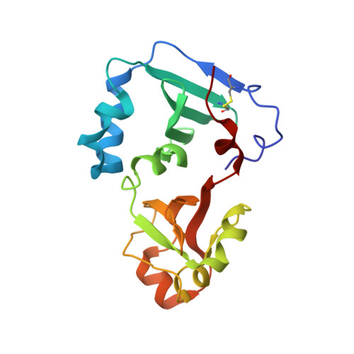
Crystal structure of the DENR-MCT-1 complex revealed zinc-binding site essential for heterodimer formation.

Lomakin, I.B.Dmitriev, S.E.Steitz, T.A.

(2019) Proc Natl Acad Sci U S A 116: 528-533

  • DOI: https://doi.org/10.1073/pnas.1809688116
  • Primary Citation Related Structures: 
    6MS4

  • PubMed Abstract: 

    The density-regulated protein (DENR) and the malignant T cell-amplified sequence 1 (MCT-1/MCTS1) oncoprotein support noncanonical translation initiation, promote translation reinitiation on a specific set of mRNAs with short upstream reading frames, and regulate ribosome recycling. DENR and MCT-1 form a heterodimer, which binds to the ribosome. We determined the crystal structure of the heterodimer formed by human MCT-1 and the N-terminal domain of DENR at 2.0-Å resolution. The structure of the heterodimer reveals atomic details of the mechanism of DENR and MCT-1 interaction. Four conserved cysteine residues of DENR (C34, C37, C44, C53) form a classical tetrahedral zinc ion-binding site, which preserves the structure of the DENR's MCT-1-binding interface that is essential for the dimerization. Substitution of all four cysteines by alanine abolished a heterodimer formation. Our findings elucidate further the mechanism of regulation of DENR-MCT-1 activities in unconventional translation initiation, reinitiation, and recycling.


  • Organizational Affiliation
    • Department of Molecular Biophysics and Biochemistry, Yale University, New Haven, CT 06520-8114; ivan.lomakin@yale.edu.

Macromolecule Content 

  • Total Structure Weight: 26.7 kDa 
  • Atom Count: 1,862 
  • Modeled Residue Count: 227 
  • Deposited Residue Count: 227 
  • Unique protein chains: 2

Macromolecules

Find similar proteins by:|  3D Structure
Entity ID: 1
MoleculeChains  Sequence LengthOrganismDetailsImage
Malignant T-cell-amplified sequence 1181Homo sapiensMutation(s): 0 
Gene Names: MCTS1MCT1
UniProt & NIH Common Fund Data Resources
Find proteins for Q9ULC4 (Homo sapiens)
Explore Q9ULC4 
Go to UniProtKB:  Q9ULC4
PHAROS:  Q9ULC4
GTEx:  ENSG00000232119 
Entity Groups
Sequence Clusters30% Identity50% Identity70% Identity90% Identity95% Identity100% Identity
UniProt GroupQ9ULC4
Sequence Annotations
Expand
Reference Sequence
Find similar proteins by:|  3D Structure
Entity ID: 2
MoleculeChains  Sequence LengthOrganismDetailsImage
Density-regulated protein46Homo sapiensMutation(s): 1 
Gene Names: DENRDRP1H14
UniProt & NIH Common Fund Data Resources
Find proteins for O43583 (Homo sapiens)
Explore O43583 
Go to UniProtKB:  O43583
PHAROS:  O43583
GTEx:  ENSG00000139726 
Entity Groups
Sequence Clusters30% Identity50% Identity70% Identity90% Identity95% Identity100% Identity
UniProt GroupO43583
Sequence Annotations
Expand
Reference Sequence

Small Molecules

Ligands 4 Unique
IDChains Name / Formula / InChI Key2D Diagram3D Interactions
PGE

Query on PGE


Download:Ideal Coordinates CCD File
C [auth A],
D [auth A],
H [auth B]
TRIETHYLENE GLYCOL
C6 H14 O4
ZIBGPFATKBEMQZ-UHFFFAOYSA-N
GOL

Query on GOL


Download:Ideal Coordinates CCD File
E [auth A],
I [auth B]
GLYCEROL
C3 H8 O3
PEDCQBHIVMGVHV-UHFFFAOYSA-N
ZN

Query on ZN


Download:Ideal Coordinates CCD File
G [auth B]ZINC ION
Zn
PTFCDOFLOPIGGS-UHFFFAOYSA-N
NA

Query on NA


Download:Ideal Coordinates CCD File
F [auth A]SODIUM ION
Na
FKNQFGJONOIPTF-UHFFFAOYSA-N

Experimental Data & Validation

Experimental Data

  • Method: X-RAY DIFFRACTION
  • Resolution: 2.00 Å
  • R-Value Free:  0.271 (Depositor), 0.272 (DCC) 
  • R-Value Work:  0.231 (Depositor), 0.236 (DCC) 
  • R-Value Observed: 0.233 (Depositor) 
Space Group: P 41 21 2
Unit Cell:
Length ( Å )Angle ( ˚ )
a = 67.81α = 90
b = 67.81β = 90
c = 124.75γ = 90
Software Package:
Software NamePurpose
PHENIXrefinement
XDSdata reduction
XSCALEdata scaling
PDB_EXTRACTdata extraction
PHASERphasing

Structure Validation

View Full Validation Report


Entry History 

& Funding Information

Deposition Data


Funding OrganizationLocationGrant Number
Howard Hughes Medical Institute (HHMI)United States--
National Institutes of Health/National Institute of General Medical Sciences (NIH/NIGMS)United StatesGM022778

Revision History  (Full details and data files)

  • Version 1.0: 2019-01-02
    Type: Initial release
  • Version 1.1: 2019-01-09
    Changes: Data collection, Database references
  • Version 1.2: 2019-01-23
    Changes: Data collection, Database references
  • Version 1.3: 2019-11-20
    Changes: Author supporting evidence
  • Version 1.4: 2023-10-11
    Changes: Data collection, Database references, Refinement description
  • Version 1.5: 2024-11-06
    Changes: Structure summary