6MRG | pdb_00006mrg

FAAH bound to non covalent inhibitor


Experimental Data Snapshot

  • Method: X-RAY DIFFRACTION
  • Resolution: 2.77 Å
  • R-Value Free: 
    0.240 (Depositor), 0.242 (DCC) 
  • R-Value Work: 
    0.195 (Depositor), 0.194 (DCC) 
  • R-Value Observed: 
    0.192 (Depositor) 

wwPDB Validation 3D Report Full Report

Validation slider image for 6MRG

Ligand Structure Quality Assessment 


This is version 1.2 of the entry. See complete history

Literature

Predicting the Binding of Fatty Acid Amide Hydrolase Inhibitors by Free Energy Perturbation.

Saha, A.Shih, A.Y.Mirzadegan, T.Seierstad, M.

(2018) J Chem Theory Comput 14: 5815-5822

  • DOI: https://doi.org/10.1021/acs.jctc.8b00672
  • Primary Citation Related Structures: 
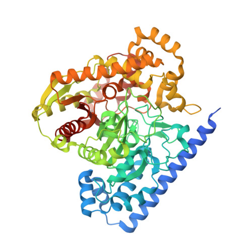
    6MRG

  • PubMed Abstract: 

    Since a goal of most drug discovery projects in either academia or industry is to design molecules that selectively bind to the desired protein, determination of protein-ligand binding free energies is of utmost importance in computer aided drug design. With the help of significant improvements in computer power, enhanced sampling techniques and accuracy of force fields, FEP (free energy perturbation) is becoming an important tool to estimate binding free energies in many drug discovery projects both retrospectively and prospectively. We have evaluated the ability of Schrödinger's FEP+ to predict relative binding free energies of a congeneric series of noncovalent fatty acid amide hydrolase (FAAH) inhibitors using an in-house crystal structure. This study shows that although an impressively accurate correlation can be obtained with experimental IC 50 s considering small perturbations on the deeper side of the pocket, the same was not observed with small perturbations on the relatively more open-ended and solvent-accessible side of the pocket. To understand these observations, we thoroughly investigated several key factors including the sampling of asymmetrically substituted rings, different perturbation maps, impact of simultaneous perturbations at two different ends of the ligand, and selecting the perturbations in a "chemically sensible" way.


  • Organizational Affiliation
    • Janssen Research and Development , 3210 Merryfield Row , San Diego , California 92121 , United States.

Macromolecule Content 

  • Total Structure Weight: 256.51 kDa 
  • Atom Count: 16,863 
  • Modeled Residue Count: 2,169 
  • Deposited Residue Count: 2,320 
  • Unique protein chains: 1

Macromolecules

Find similar proteins by:|  3D Structure
Entity ID: 1
MoleculeChains  Sequence LengthOrganismDetailsImage
Fatty-acid amide hydrolase 1
A, B, C, D
580Rattus norvegicusMutation(s): 0 
Gene Names: FaahFaah1
EC: 3.5.1.99 (PDB Primary Data), 3.1.1 (UniProt)
UniProt
Find proteins for P97612 (Rattus norvegicus)
Explore P97612 
Go to UniProtKB:  P97612
Entity Groups
Sequence Clusters30% Identity50% Identity70% Identity90% Identity95% Identity100% Identity
UniProt GroupP97612
Sequence Annotations
Expand
Reference Sequence

Experimental Data & Validation

Experimental Data

  • Method: X-RAY DIFFRACTION
  • Resolution: 2.77 Å
  • R-Value Free:  0.240 (Depositor), 0.242 (DCC) 
  • R-Value Work:  0.195 (Depositor), 0.194 (DCC) 
  • R-Value Observed: 0.192 (Depositor) 
Space Group: C 2 2 21
Unit Cell:
Length ( Å )Angle ( ˚ )
a = 144.208α = 90
b = 152.014β = 90
c = 299.914γ = 90
Software Package:
Software NamePurpose
PHENIXrefinement
XDSdata reduction
XSCALEdata scaling
MOLREPphasing

Structure Validation

View Full Validation Report


Ligand Structure Quality Assessment 


Entry History 

Deposition Data

Revision History  (Full details and data files)

  • Version 1.0: 2018-10-31
    Type: Initial release
  • Version 1.1: 2018-11-21
    Changes: Data collection, Database references
  • Version 1.2: 2024-03-13
    Changes: Advisory, Data collection, Database references