6MKZ | pdb_00006mkz

Crystal structure of murine 4-1BB/4-1BBL complex


Experimental Data Snapshot

  • Method: X-RAY DIFFRACTION
  • Resolution: 2.65 Å
  • R-Value Free: 
    0.238 (Depositor), 0.252 (DCC) 
  • R-Value Work: 
    0.204 (Depositor), 0.224 (DCC) 
  • R-Value Observed: 
    0.206 (Depositor) 

Starting Models: experimental
View more details

wwPDB Validation 3D Report Full Report

Validation slider image for 6MKZ

This is version 2.2 of the entry. See complete history

Literature

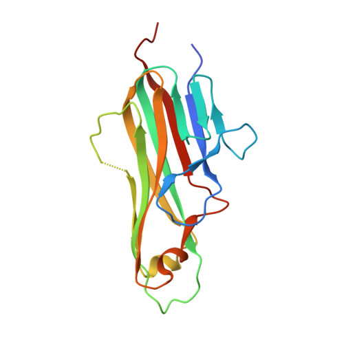
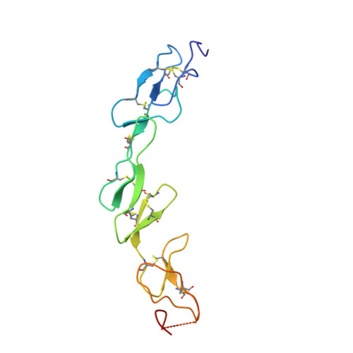
Crystal structure of the m4-1BB/4-1BBL complex reveals an unusual dimeric ligand that undergoes structural changes upon 4-1BB receptor binding.

Bitra, A.Doukov, T.Destito, G.Croft, M.Zajonc, D.M.

(2019) J Biological Chem 294: 1831-1845

  • DOI: https://doi.org/10.1074/jbc.RA118.006297
  • Primary Citation Related Structures: 
    6MKB, 6MKZ

  • PubMed Abstract: 

    The interaction between the receptor 4-1BB and its ligand 4-1BBL provides co-stimulatory signals for T-cell activation and proliferation. However, differences in the mouse and human molecules might result in differential engagement of this pathway. Here, we report the crystal structure of mouse 4-1BBL and of the mouse 4-1BB/4-1BBL complex, which together provided insights into the molecular mechanism by which m4-1BBL and its cognate receptor recognize each other. Unlike all human or mouse tumor necrosis factor ligands that form noncovalent and mostly trimeric assemblies, the m4-1BBL structure formed a disulfide-linked dimeric assembly. The structure disclosed that certain differences in the amino acid composition along the intramolecular interface, together with two specific residues (Cys-246 and Ser-256) present exclusively in m4-1BBL, are responsible for this unique dimerization. Unexpectedly, upon m4-1BB binding, m4-1BBL undergoes structural changes within each protomer; moreover, the individual m4-1BBL protomers rotate relative to each other, yielding a dimerization interface with more inter-subunit interactions. We also observed that in the m4-1BB/4-1BBL complex, each receptor monomer binds exclusively to a single ligand subunit with contributions of cysteine-rich domain 1 (CRD1), CRD2, and CRD3. Furthermore, structure-guided mutagenesis of the binding interface revealed that novel binding interactions with the GH loop, rather than the DE loop, are energetically critical and define the m4-1BB receptor selectivity for m4-1BBL. A comparison with the human 4-1BB/4-1BBL complex highlighted several differences between the ligand- and receptor-binding interfaces, providing an explanation for the absence of inter-species cross-reactivity between human and mouse 4-1BB and 4-1BBL molecules.


  • Organizational Affiliation
    • From the Division of Immune Regulation, La Jolla Institute for Immunology (LJI), La Jolla, California 92037.

Macromolecule Content 

  • Total Structure Weight: 69.98 kDa 
  • Atom Count: 4,179 
  • Modeled Residue Count: 528 
  • Deposited Residue Count: 620 
  • Unique protein chains: 2

Macromolecules

Find similar proteins by:|  3D Structure
Entity ID: 1
MoleculeChains  Sequence LengthOrganismDetailsImage
Tumor necrosis factor ligand superfamily member 9
A, C
171Mus musculusMutation(s): 0 
Gene Names: Tnfsf9Cd137lCd157lLy63l
UniProt
Find proteins for P41274 (Mus musculus)
Explore P41274 
Go to UniProtKB:  P41274
Entity Groups
Sequence Clusters30% Identity50% Identity70% Identity90% Identity95% Identity100% Identity
UniProt GroupP41274
Glycosylation
Glycosylation Sites: 1Go to GlyGen: P41274-1
Sequence Annotations
Expand
Reference Sequence
Find similar proteins by:|  3D Structure
Entity ID: 2
MoleculeChains  Sequence LengthOrganismDetailsImage
Tumor necrosis factor receptor superfamily member 9
B, D
139Mus musculusMutation(s): 0 
Gene Names: Tnfrsf9Cd137IlaLy63
UniProt & NIH Common Fund Data Resources
Find proteins for P20334 (Mus musculus)
Explore P20334 
Go to UniProtKB:  P20334
IMPC:  MGI:1101059
Entity Groups
Sequence Clusters30% Identity50% Identity70% Identity90% Identity95% Identity100% Identity
UniProt GroupP20334
Sequence Annotations
Expand
Reference Sequence

Oligosaccharides

Help  
Entity ID: 3
MoleculeChains Length2D Diagram GlycosylationD Interactions
beta-D-mannopyranose-(1-4)-2-acetamido-2-deoxy-beta-D-glucopyranose-(1-4)-2-acetamido-2-deoxy-beta-D-glucopyranose
E, F
3N-Glycosylation
Glycosylation Resources
GlyTouCan: G15407YE
GlyCosmos: G15407YE
GlyGen: G15407YE

Experimental Data & Validation

Experimental Data

  • Method: X-RAY DIFFRACTION
  • Resolution: 2.65 Å
  • R-Value Free:  0.238 (Depositor), 0.252 (DCC) 
  • R-Value Work:  0.204 (Depositor), 0.224 (DCC) 
  • R-Value Observed: 0.206 (Depositor) 
Space Group: P 21 21 21
Unit Cell:
Length ( Å )Angle ( ˚ )
a = 61.499α = 90
b = 88.478β = 90
c = 155.158γ = 90
Software Package:
Software NamePurpose
BUSTERrefinement
Aimlessdata scaling
PDB_EXTRACTdata extraction
XDSdata reduction
PHASERphasing

Structure Validation

View Full Validation Report


Entry History 

Deposition Data

Revision History  (Full details and data files)

  • Version 1.0: 2018-12-19
    Type: Initial release
  • Version 1.1: 2018-12-26
    Changes: Data collection, Database references
  • Version 1.2: 2019-02-20
    Changes: Data collection, Database references
  • Version 2.0: 2020-07-29
    Type: Remediation
    Reason: Carbohydrate remediation
    Changes: Atomic model, Data collection, Derived calculations, Structure summary
  • Version 2.1: 2023-10-11
    Changes: Data collection, Database references, Refinement description, Structure summary
  • Version 2.2: 2024-11-06
    Changes: Structure summary