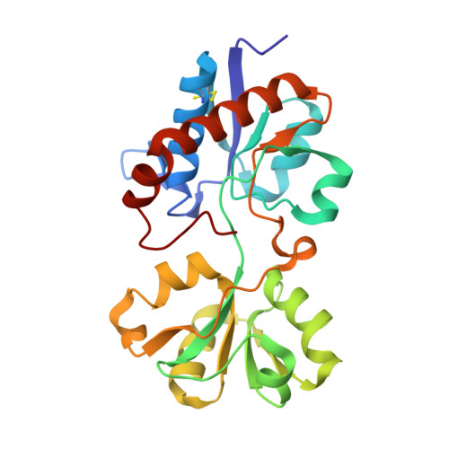
The interplay of protein-ligand and water-mediated interactions shape affinity and selectivity in the LAO binding protein.
Vergara, R., Romero-Romero, S., Velazquez-Lopez, I., Espinoza-Perez, G., Rodriguez-Hernandez, A., Pulido, N.O., Sosa-Peinado, A., Rodriguez-Romero, A., Fernandez-Velasco, D.A.(2020) FEBS J 287: 763-782
- PubMed: 31348608
- DOI: https://doi.org/10.1111/febs.15019
- Primary Citation of Related Structures:
6MKU, 6MKW, 6MKX, 6ML0, 6ML9, 6MLA, 6MLD, 6MLE, 6MLG, 6MLI, 6MLJ, 6MLN, 6MLO, 6MLP, 6MLV - PubMed Abstract:
The study of binding thermodynamics is essential to understand how affinity and selectivity are acquired in molecular complexes. Periplasmic binding proteins (PBPs) are macromolecules of biotechnological interest that bind a broad number of ligands and have been used to design biosensors. The lysine-arginine-ornithine binding protein (LAO) is a PBP of 238 residues that binds the basic amino acids l-arginine and l-histidine with nm and μm affinity, respectively. It has been shown that the affinity difference for arginine and histidine binding is caused by enthalpy, this correlates with the higher number of protein-ligand contacts formed with arginine. In order to elucidate the structural bases that determine binding affinity and selectivity in LAO, the contribution of protein-ligand contacts to binding energetics was assessed. To this end, an alanine scanning of the LAO-binding site residues was performed and arginine and histidine binding were characterized by isothermal titration calorimetry and X-ray crystallography. Although unexpected enthalpy and entropy changes were observed in some mutants, thermodynamic data correlated with structural information, especially, the binding heat capacity change. We found that selectivity is conferred by several residues rather than exclusive arginine-protein interactions. Furthermore, crystallographic structures revealed that protein-ligand contributions to binding thermodynamics are highly influenced by the solvent. Finally, we found a similar backbone conformation in all the closed structures obtained, but different structures in the open state, suggesting that the binding site residues of LAO play an important role in stabilizing not only the holo conformation, but also the apo state. DATABASE: Structural data are available in the Protein Data Bank database under the accession numbers 6MLE, 6MLN, 6MLG, 6MKX, 6MLI, 6MLA, 6MKU, 6MKW, 6ML0, 6MLD, 6MLV, 6MLO, 6MLP, 6ML9, 6MLJ.
- Laboratorio de Fisicoquímica e Ingeniería de Proteínas, Departamento de Bioquímica, Facultad de Medicina, Universidad Nacional Autónoma de México, Ciudad de México, México.
Organizational Affiliation:


