6MJH | pdb_00006mjh

The S31N mutant of the influenza A M2 proton channel in two distinct conformational states


Experimental Data Snapshot

  • Method: X-RAY DIFFRACTION
  • Resolution: 2.06 Å
  • R-Value Free: 
    0.248 (Depositor), 0.251 (DCC) 
  • R-Value Work: 
    0.217 (Depositor), 0.218 (DCC) 
  • R-Value Observed: 
    0.220 (Depositor) 

Starting Models: experimental
View more details

wwPDB Validation 3D Report Full Report

Validation slider image for 6MJH

This is version 1.4 of the entry. See complete history

Literature

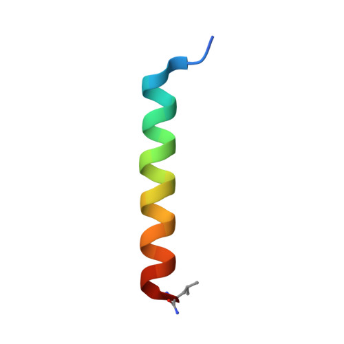
X-ray Crystal Structure of the Influenza A M2 Proton Channel S31N Mutant in Two Conformational States: An Open and Shut Case.

Thomaston, J.L.Wu, Y.Polizzi, N.Liu, L.Wang, J.DeGrado, W.F.

(2019) J Am Chem Soc 141: 11481-11488

  • DOI: https://doi.org/10.1021/jacs.9b02196
  • Primary Citation Related Structures: 
    6MJH

  • PubMed Abstract: 

    The amantadine-resistant S31N mutant of the influenza A M2 proton channel has become prevalent in currently circulating viruses. Here, we have solved an X-ray crystal structure of M2(22-46) S31N that contains two distinct conformational states within its asymmetric unit. This structure reveals the mechanism of adamantane resistance in both conformational states of the M2 channel. In the Inward open conformation, the mutant Asn31 side chain faces the channel pore and sterically blocks the adamantane binding site. In the Inward closed conformation, Asn31 forms hydrogen bonds with carbonyls at the monomer-monomer interface, which twists the monomer helices and constricts the channel pore at the drug binding site. We also examine M2(19-49) WT and S31N using solution NMR spectroscopy and show that distribution of the two conformational states is dependent on both detergent choice and experimental pH.


  • Organizational Affiliation
    • Department of Pharmaceutical Chemistry , University of California , San Francisco , California 94158 , United States.

Macromolecule Content 

  • Total Structure Weight: 22.45 kDa 
  • Atom Count: 1,666 
  • Modeled Residue Count: 216 
  • Deposited Residue Count: 216 
  • Unique protein chains: 1

Macromolecules

Find similar proteins by:|  3D Structure
Entity ID: 1
MoleculeChains  Sequence LengthOrganismDetailsImage
Matrix protein 2
A, B, C, D, E
A, B, C, D, E, F, G, H
27Influenza A virus (A/pigeon/Jiangsu/K23/2013(H9N2))Mutation(s): 2 
Membrane Entity: Yes 
UniProt
Find proteins for A0A0R5TVW3 (Influenza A virus)
Explore A0A0R5TVW3 
Go to UniProtKB:  A0A0R5TVW3
Entity Groups
Sequence Clusters30% Identity50% Identity70% Identity90% Identity95% Identity100% Identity
UniProt GroupA0A0R5TVW3
Sequence Annotations
Expand
Reference Sequence

Experimental Data & Validation

Experimental Data

  • Method: X-RAY DIFFRACTION
  • Resolution: 2.06 Å
  • R-Value Free:  0.248 (Depositor), 0.251 (DCC) 
  • R-Value Work:  0.217 (Depositor), 0.218 (DCC) 
  • R-Value Observed: 0.220 (Depositor) 
Space Group: P 1 21 1
Unit Cell:
Length ( Å )Angle ( ˚ )
a = 36.29α = 90
b = 36.15β = 103.6
c = 76.45γ = 90
Software Package:
Software NamePurpose
PHENIXrefinement
iMOSFLMdata reduction
Aimlessdata scaling
PHASERphasing

Structure Validation

View Full Validation Report


Entry History 

& Funding Information

Deposition Data


Funding OrganizationLocationGrant Number
National Institutes of Health/National Human Genome Research Institute (NIH/NHGRI)United States--

Revision History  (Full details and data files)

  • Version 1.0: 2019-06-26
    Type: Initial release
  • Version 1.1: 2019-08-07
    Changes: Data collection, Database references
  • Version 1.2: 2019-12-18
    Changes: Author supporting evidence
  • Version 1.3: 2023-10-11
    Changes: Data collection, Database references, Derived calculations, Refinement description
  • Version 1.4: 2024-11-06
    Changes: Structure summary