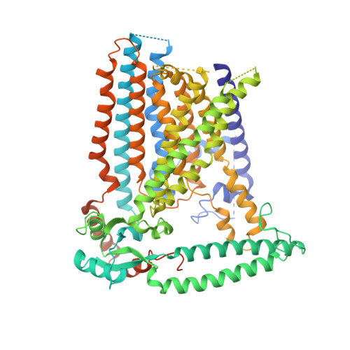
6MGW

Structure of mechanically activated ion channel OSCA1.2 in LMNG


Experimental Data Snapshot

  • Method: ELECTRON MICROSCOPY
  • Resolution: 3.50 Å
  • Aggregation State: PARTICLE 
  • Reconstruction Method: SINGLE PARTICLE 

wwPDB Validation   3D Report Full Report


This is version 1.3 of the entry. See complete history


Literature

Cryo-EM structure of the mechanically activated ion channel OSCA1.2.

Jojoa Cruz, S.Saotome, K.Murthy, S.E.Tsui, C.C.A.Sansom, M.S.Patapoutian, A.Ward, A.B.

(2018) Elife 7

  • DOI: https://doi.org/10.7554/eLife.41845
  • Primary Citation of Related Structures:  
    6MGV, 6MGW

  • PubMed Abstract: 

    Mechanically activated ion channels underlie touch, hearing, shear-stress sensing, and response to turgor pressure. OSCA/TMEM63s are a newly-identified family of eukaryotic mechanically activated ion channels opened by membrane tension. The structural underpinnings of OSCA/TMEM63 function are not explored. Here, we elucidate high resolution cryo-electron microscopy structures of OSCA1.2, revealing a dimeric architecture containing eleven transmembrane helices per subunit and surprising topological similarities to TMEM16 proteins. We locate the ion permeation pathway within each subunit by demonstrating that a conserved acidic residue is a determinant of channel conductance. Molecular dynamics simulations reveal membrane interactions, suggesting the role of lipids in OSCA1.2 gating. These results lay a foundation to decipher how the structural organization of OSCA/TMEM63 is suited for their roles as MA ion channels.


  • Organizational Affiliation

    Department of Integrative Structural and Computational Biology, The Scripps Research Institute, California, United States.


Macromolecules
Find similar proteins by:  (by identity cutoff)  |  3D Structure
Entity ID: 1
MoleculeChains Sequence LengthOrganismDetailsImage
Calcium permeable stress-gated cation channel 1
A, B
781Arabidopsis thalianaMutation(s): 0 
Gene Names: CSC1At4g22120F1N20.220
Membrane Entity: Yes 
UniProt
Find proteins for Q5XEZ5 (Arabidopsis thaliana)
Explore Q5XEZ5 
Go to UniProtKB:  Q5XEZ5
Entity Groups  
Sequence Clusters30% Identity50% Identity70% Identity90% Identity95% Identity100% Identity
UniProt GroupQ5XEZ5
Sequence Annotations
Expand
  • Reference Sequence
Experimental Data & Validation

Experimental Data

  • Method: ELECTRON MICROSCOPY
  • Resolution: 3.50 Å
  • Aggregation State: PARTICLE 
  • Reconstruction Method: SINGLE PARTICLE 
EM Software:
TaskSoftware PackageVersion
RECONSTRUCTIONcryoSPARC

Structure Validation

View Full Validation Report


Entry History & Funding Information

Deposition Data


Funding OrganizationLocationGrant Number
Howard Hughes Medical Institute (HHMI)United States--
National Institutes of Health/National Institute of Neurological Disorders and Stroke (NIH/NINDS)United States1R35NS105067

Revision History  (Full details and data files)

  • Version 1.0: 2018-11-14
    Type: Initial release
  • Version 1.1: 2019-11-06
    Changes: Author supporting evidence, Data collection
  • Version 1.2: 2019-11-20
    Changes: Author supporting evidence
  • Version 1.3: 2024-03-13
    Changes: Data collection, Database references