6MF8

TCR alpha transmembrane domain

  • Classification: IMMUNE SYSTEM
  • Organism(s): Mus musculus
  • Expression System: Escherichia coli
  • Mutation(s): No 
  • Membrane Protein: Yes  OPMPDBTM

  • Deposited: 2018-09-10 Released: 2018-12-12 
  • Deposition Author(s): Brazin, K.N., Reinherz, E.L.
  • Funding Organization(s): National Institutes of Health/National Institute of General Medical Sciences (NIH/NIGMS), National Institutes of Health/National Institute Of Allergy and Infectious Diseases (NIH/NIAID)

Experimental Data Snapshot

  • Method: SOLUTION NMR
  • Conformers Calculated: 100 
  • Conformers Submitted: 10 
  • Selection Criteria: structures with the lowest energy 

wwPDB Validation   3D Report Full Report


This is version 1.1 of the entry. See complete history


Literature

The T Cell Antigen Receptor alpha Transmembrane Domain Coordinates Triggering through Regulation of Bilayer Immersion and CD3 Subunit Associations.

Brazin, K.N.Mallis, R.J.Boeszoermenyi, A.Feng, Y.Yoshizawa, A.Reche, P.A.Kaur, P.Bi, K.Hussey, R.E.Duke-Cohan, J.S.Song, L.Wagner, G.Arthanari, H.Lang, M.J.Reinherz, E.L.

(2018) Immunity 49: 829-841.e6

  • DOI: https://doi.org/10.1016/j.immuni.2018.09.007
  • Primary Citation of Related Structures:  
    6MF8

  • PubMed Abstract: 

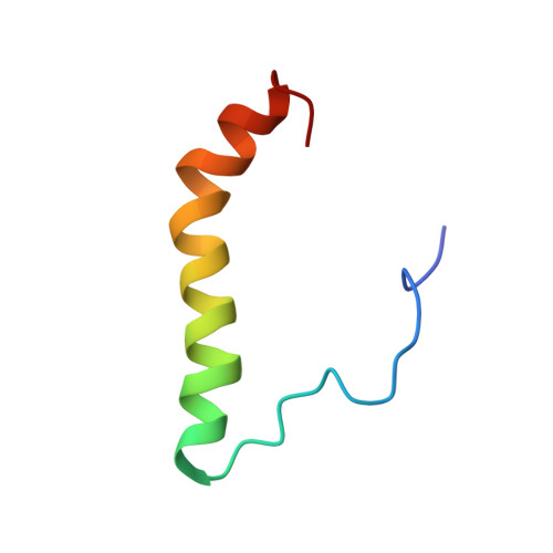
    Initial molecular details of cellular activation following αβT cell antigen receptor (TCR) ligation by peptide-major histocompatibility complexes (pMHC) remain unexplored. We determined the nuclear magnetic resonance (NMR) structure of the TCRα subunit transmembrane (TM) domain revealing a bipartite helix whose segmentation fosters dynamic movement. Positively charged TM residues Arg251 and Lys256 project from opposite faces of the helix, with Lys256 controlling immersion depth. Their modification caused stepwise reduction in TCR associations with CD3ζζ homodimers and CD3εγ plus CD3εδ heterodimers, respectively, leading to an activated transcriptome. Optical tweezers revealed that Arg251 and Lys256 mutations altered αβTCR-pMHC bond lifetimes, while mutations within interacting TCRα connecting peptide and CD3δ CxxC motif juxtamembrane elements selectively attenuated signal transduction. Our findings suggest that mechanical forces applied during pMHC ligation initiate T cell activation via a dissociative mechanism, shifting disposition of those basic sidechains to rearrange TCR complex membrane topology and weaken TCRαβ and CD3 associations.


  • Organizational Affiliation

    Laboratory of Immunobiology and Department of Medical Oncology, Dana-Farber Cancer Institute, and Department of Medicine, Harvard Medical School, Boston, MA 02115, USA.


Macromolecules
Find similar proteins by:  (by identity cutoff)  |  3D Structure
Entity ID: 1
MoleculeChains Sequence LengthOrganismDetailsImage
T-cell receptor alpha chain C region46Mus musculusMutation(s): 0 
Gene Names: Tcra
Membrane Entity: Yes 
UniProt
Find proteins for P01849 (Mus musculus)
Explore P01849 
Go to UniProtKB:  P01849
Entity Groups  
Sequence Clusters30% Identity50% Identity70% Identity90% Identity95% Identity100% Identity
UniProt GroupP01849
Sequence Annotations
Expand
  • Reference Sequence
Experimental Data & Validation

Experimental Data

  • Method: SOLUTION NMR
  • Conformers Calculated: 100 
  • Conformers Submitted: 10 
  • Selection Criteria: structures with the lowest energy 

Structure Validation

View Full Validation Report


Entry History & Funding Information

Deposition Data


Funding OrganizationLocationGrant Number
National Institutes of Health/National Institute of General Medical Sciences (NIH/NIGMS)United StatesGM047467
National Institutes of Health/National Institute Of Allergy and Infectious Diseases (NIH/NIAID)United StatesRO1AI100643
National Institutes of Health/National Institute Of Allergy and Infectious Diseases (NIH/NIAID)United StatesR56AI138489

Revision History  (Full details and data files)

  • Version 1.0: 2018-12-12
    Type: Initial release
  • Version 1.1: 2019-12-18
    Changes: Author supporting evidence, Data collection