6ME1

Crystal structure of the clade B isolate B41 mutant fusion peptide (residues 512-521) in complex with VRC34.01


Experimental Data Snapshot

  • Method: X-RAY DIFFRACTION
  • Resolution: 1.97 Å
  • R-Value Free: 0.238 
  • R-Value Work: 0.199 
  • R-Value Observed: 0.201 

wwPDB Validation   3D Report Full Report

Currently 6ME1 does not have a validation slider image.


This is version 1.1 of the entry. See complete history


Literature

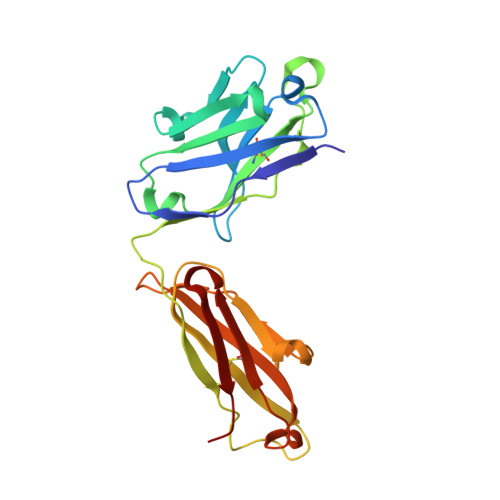
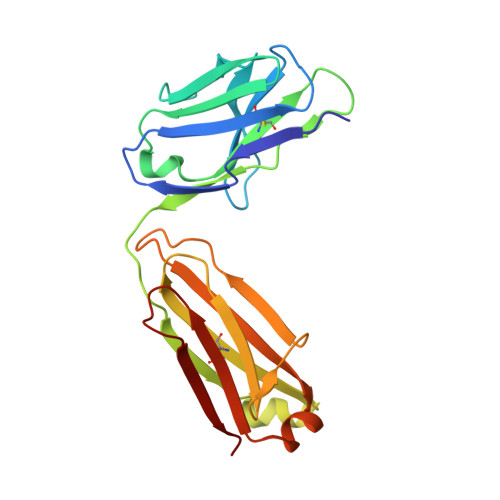
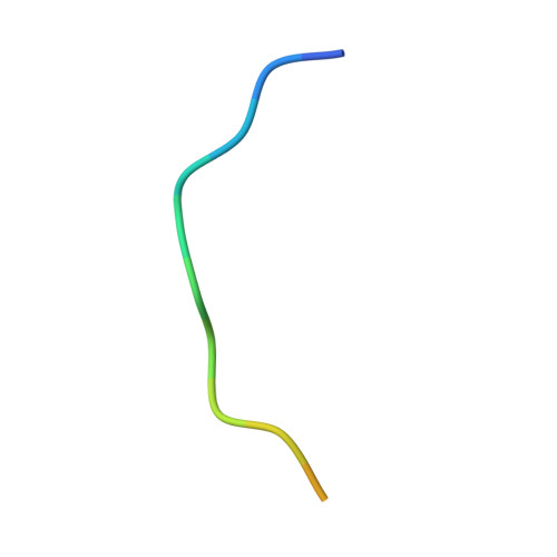
Capturing the inherent structural dynamics of the HIV-1 envelope glycoprotein fusion peptide.

Kumar, S.Sarkar, A.Pugach, P.Sanders, R.W.Moore, J.P.Ward, A.B.Wilson, I.A.

(2019) Nat Commun 10: 763-763

  • DOI: https://doi.org/10.1038/s41467-019-08738-5
  • Primary Citation of Related Structures:  
    6MCO, 6MDT, 6ME1

  • PubMed Abstract: 

    The N-terminal fusion peptide (FP) of the human immunodeficiency virus (HIV)-1 envelope glycoprotein (Env) gp41 subunit plays a critical role in cell entry. However, capturing the structural flexibility in the unbound FP is challenging in the native Env trimer. Here, FP conformational isomerism is observed in two crystal structures of a soluble clade B transmitted/founder virus B41 SOSIP.664 Env with broadly neutralizing antibodies (bNAbs) PGT124 and 35O22 to aid in crystallization and that are not specific for binding to the FP. Large rearrangements in the FP and fusion peptide proximal region occur around M530, which remains anchored in the tryptophan clasp (gp41 W623, W628, W631) in the B41 Env prefusion state. Further, we redesigned the FP at position 518 to reinstate the bNAb VRC34.01 epitope. These findings provide further structural evidence for the dynamic nature of the FP and how a bNAb epitope can be restored during vaccine design.


  • Organizational Affiliation

    Department of Integrative Structural and Computational Biology, The Scripps Research Institute, La Jolla, CA, 92037, USA.


Macromolecules
Find similar proteins by:  (by identity cutoff)  |  3D Structure
Entity ID: 1
MoleculeChains Sequence LengthOrganismDetailsImage
VRC34.01 Fab heavy chainA,
D [auth C]
223Homo sapiensMutation(s): 0 
Entity Groups  
Sequence Clusters30% Identity50% Identity70% Identity90% Identity95% Identity100% Identity
Sequence Annotations
Expand
  • Reference Sequence
Find similar proteins by:  (by identity cutoff)  |  3D Structure
Entity ID: 2
MoleculeChains Sequence LengthOrganismDetailsImage
VRC34.01 Fab light chainB,
E [auth D]
212Homo sapiensMutation(s): 0 
Entity Groups  
Sequence Clusters30% Identity50% Identity70% Identity90% Identity95% Identity100% Identity
Sequence Annotations
Expand
  • Reference Sequence

Find similar proteins by:  Sequence   |   3D Structure  

Entity ID: 3
MoleculeChains Sequence LengthOrganismDetailsImage
Envelope glycoprotein gp160C [auth F],
F [auth E]
16Human immunodeficiency virus 1Mutation(s): 0 
UniProt
Find proteins for P03375 (Human immunodeficiency virus type 1 group M subtype B (isolate BH10))
Explore P03375 
Go to UniProtKB:  P03375
Entity Groups  
Sequence Clusters30% Identity50% Identity70% Identity90% Identity95% Identity100% Identity
UniProt GroupP03375
Sequence Annotations
Expand
  • Reference Sequence
Experimental Data & Validation

Experimental Data

  • Method: X-RAY DIFFRACTION
  • Resolution: 1.97 Å
  • R-Value Free: 0.238 
  • R-Value Work: 0.199 
  • R-Value Observed: 0.201 
  • Space Group: P 1 21 1
Unit Cell:
Length ( Å )Angle ( ˚ )
a = 42.421α = 90
b = 123.67β = 90.87
c = 101.253γ = 90
Software Package:
Software NamePurpose
PHENIXrefinement
PHASERphasing
HKL-2000data scaling
HKL-2000data reduction

Structure Validation

View Full Validation Report

Currently 6ME1 does not have a validation slider image.


Entry History 

Deposition Data

Revision History  (Full details and data files)

  • Version 1.0: 2019-02-27
    Type: Initial release
  • Version 1.1: 2023-10-11
    Changes: Data collection, Database references, Refinement description