6MC9 | pdb_00006mc9

Crystal Structure of Human Nav1.4 C-Terminal (1599-1754) domain in complex with calcium-bound calmodulin


Experimental Data Snapshot

  • Method: X-RAY DIFFRACTION
  • Resolution: 3.30 Å
  • R-Value Free: 
    0.285 (Depositor), 0.285 (DCC) 
  • R-Value Work: 
    0.240 (Depositor), 0.242 (DCC) 
  • R-Value Observed: 
    0.242 (Depositor) 

Starting Model: experimental
View more details

wwPDB Validation 3D Report Full Report

Validation slider image for 6MC9

This is version 1.3 of the entry. See complete history

Literature

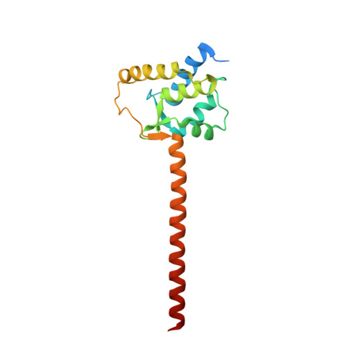
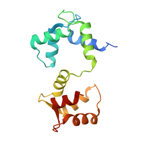
Ca2+-dependent regulation of sodium channels NaV1.4 and NaV1.5 is controlled by the post-IQ motif.

Yoder, J.B.Ben-Johny, M.Farinelli, F.Srinivasan, L.Shoemaker, S.R.Tomaselli, G.F.Gabelli, S.B.Amzel, L.M.

(2019) Nat Commun 10: 1514-1514

  • DOI: https://doi.org/10.1038/s41467-019-09570-7
  • Primary Citation Related Structures: 
    6MBA, 6MC9

  • PubMed Abstract: 

    Skeletal muscle voltage-gated Na + channel (Na V 1.4) activity is subject to calmodulin (CaM) mediated Ca 2+ -dependent inactivation; no such inactivation is observed in the cardiac Na + channel (Na V 1.5). Taken together, the crystal structures of the Na V 1.4 C-terminal domain relevant complexes and thermodynamic binding data presented here provide a rationale for this isoform difference. A Ca 2+ -dependent CaM N-lobe binding site previously identified in Na V 1.5 is not present in Na V 1.4 allowing the N-lobe to signal other regions of the Na V 1.4 channel. Consistent with this mechanism, removing this binding site in Na V 1.5 unveils robust Ca 2+ -dependent inactivation in the previously insensitive isoform. These findings suggest that Ca 2+ -dependent inactivation is effected by CaM's N-lobe binding outside the Na V C-terminal while CaM's C-lobe remains bound to the Na V C-terminal. As the N-lobe binding motif of Na V 1.5 is a mutational hotspot for inherited arrhythmias, the contributions of mutation-induced changes in CDI to arrhythmia generation is an intriguing possibility.


  • Organizational Affiliation
    • Department of Biophysics and Biophysical Chemistry, Johns Hopkins University School of Medicine, Baltimore, MD, 21205, USA.

Macromolecule Content 

  • Total Structure Weight: 36.22 kDa 
  • Atom Count: 2,330 
  • Modeled Residue Count: 291 
  • Deposited Residue Count: 317 
  • Unique protein chains: 2

Macromolecules

Find similar proteins by:|  3D Structure
Entity ID: 1
MoleculeChains  Sequence LengthOrganismDetailsImage
Sodium channel protein type 4 subunit alpha168Homo sapiensMutation(s): 0 
Gene Names: SCN4A
Membrane Entity: Yes 
UniProt & NIH Common Fund Data Resources
Find proteins for P35499 (Homo sapiens)
Explore P35499 
Go to UniProtKB:  P35499
PHAROS:  P35499
GTEx:  ENSG00000007314 
Entity Groups
Sequence Clusters30% Identity50% Identity70% Identity90% Identity95% Identity100% Identity
UniProt GroupP35499
Sequence Annotations
Expand
Reference Sequence
Find similar proteins by:|  3D Structure
Entity ID: 2
MoleculeChains  Sequence LengthOrganismDetailsImage
Calmodulin-1149Rattus norvegicusMutation(s): 0 
Gene Names: Calm1CalmCamCam1CaMI
UniProt
Find proteins for P0DP29 (Rattus norvegicus)
Explore P0DP29 
Go to UniProtKB:  P0DP29
Entity Groups
Sequence Clusters30% Identity50% Identity70% Identity90% Identity95% Identity100% Identity
UniProt GroupP0DP29
Sequence Annotations
Expand
Reference Sequence

Experimental Data & Validation

Experimental Data

  • Method: X-RAY DIFFRACTION
  • Resolution: 3.30 Å
  • R-Value Free:  0.285 (Depositor), 0.285 (DCC) 
  • R-Value Work:  0.240 (Depositor), 0.242 (DCC) 
  • R-Value Observed: 0.242 (Depositor) 
Space Group: P 43 21 2
Unit Cell:
Length ( Å )Angle ( ˚ )
a = 72.615α = 90
b = 72.615β = 90
c = 134.661γ = 90
Software Package:
Software NamePurpose
PHENIXrefinement
XDSdata reduction
XSCALEdata scaling
PDB_EXTRACTdata extraction
PHASERphasing

Structure Validation

View Full Validation Report


Entry History 

& Funding Information

Deposition Data


Funding OrganizationLocationGrant Number
National Institutes of Health/National Heart, Lung, and Blood Institute (NIH/NHLBI)United States5R01HL128743

Revision History  (Full details and data files)

  • Version 1.0: 2019-04-10
    Type: Initial release
  • Version 1.1: 2019-04-17
    Changes: Data collection, Database references
  • Version 1.2: 2019-12-04
    Changes: Author supporting evidence
  • Version 1.3: 2023-10-11
    Changes: Data collection, Database references, Refinement description