6MC4

Crystal structure of a tetrameric DNA fold-back quadruplex

  • Classification: DNA
  • Organism(s): synthetic construct
  • Mutation(s): No 

  • Deposited: 2018-08-30 Released: 2018-11-14 
  • Deposition Author(s): Chu, B., Paukstelis, P.J.
  • Funding Organization(s): National Science Foundation (NSF, United States)

Experimental Data Snapshot

  • Method: X-RAY DIFFRACTION
  • Resolution: 2.25 Å
  • R-Value Free: 0.268 
  • R-Value Work: 0.228 
  • R-Value Observed: 0.230 

wwPDB Validation   3D Report Full Report


This is version 1.3 of the entry. See complete history


Literature

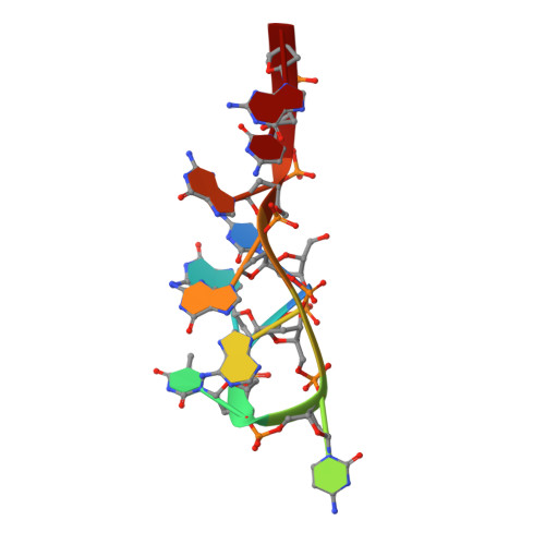
Crystal Structure of a Tetrameric DNA Fold-Back Quadruplex.

Chu, B.Zhang, D.Hwang, W.Paukstelis, P.J.

(2018) J Am Chem Soc 140: 16291-16298

  • DOI: https://doi.org/10.1021/jacs.8b10153
  • Primary Citation of Related Structures:  
    6MC2, 6MC3, 6MC4, 6N4G

  • PubMed Abstract: 

    DNA can adopt many structures beyond the Watson-Crick duplex. However, the bounds of DNA structural diversity and how these structures might regulate biological processes is only beginning to be understood. Here, we describe the 1.05 Å resolution crystal structure of a DNA oligonucleotide that self-associates to form a non-G-quadruplex fold-back structure. Distinct from previously described fold-back quadruplexes, two-fold-back dimers interact through noncanonical and Watson-Crick interactions to form a tetrameric assembly. These interactions include a hexad base pairing arrangement from two C-G-G base triples. The assembly is dependent on divalent cations, and the interface between the dimeric units creates a cavity in which a cation resides. This structure provides new sequence and structural contexts for the formation of fold-back quadruplexes, further highlighting the potential biological importance of this type of noncanonical DNA structure. This structure may also serve as the basis for designing new types of DNA nanoarchitectures or cation sensors based on the strong divalent cation dependence.


  • Organizational Affiliation

    Department of Chemistry and Biochemistry, Center for Biomolecular Structure and Organization , University of Maryland , College Park , Maryland 20742 , United States.


Macromolecules

Find similar nucleic acids by:  Sequence   |   3D Structure  

Entity ID: 1
MoleculeChains LengthOrganismImage
DNA (5'-D(*CP*GP*TP*CP*AP*GP*GP*CP*G)-3')
A, B, C, D, E
A, B, C, D, E, F, G, H
9synthetic construct
Sequence Annotations
Expand
  • Reference Sequence
Small Molecules
Ligands 2 Unique
IDChains Name / Formula / InChI Key2D Diagram3D Interactions
CA
Query on CA

Download Ideal Coordinates CCD File 
I [auth A]
K [auth C]
L [auth C]
M [auth C]
N [auth C]
I [auth A],
K [auth C],
L [auth C],
M [auth C],
N [auth C],
O [auth D],
P [auth E],
Q [auth F],
R [auth F],
S [auth H]
CALCIUM ION
Ca
BHPQYMZQTOCNFJ-UHFFFAOYSA-N
K
Query on K

Download Ideal Coordinates CCD File 
J [auth B]POTASSIUM ION
K
NPYPAHLBTDXSSS-UHFFFAOYSA-N
Experimental Data & Validation

Experimental Data

  • Method: X-RAY DIFFRACTION
  • Resolution: 2.25 Å
  • R-Value Free: 0.268 
  • R-Value Work: 0.228 
  • R-Value Observed: 0.230 
  • Space Group: P 31 2 1
Unit Cell:
Length ( Å )Angle ( ˚ )
a = 53.743α = 90
b = 53.743β = 90
c = 99.389γ = 120
Software Package:
Software NamePurpose
REFMACrefinement
XDSdata reduction
Aimlessdata scaling
PHASERphasing

Structure Validation

View Full Validation Report


Entry History & Funding Information

Deposition Data


Funding OrganizationLocationGrant Number
National Science Foundation (NSF, United States)United States1149665

Revision History  (Full details and data files)

  • Version 1.0: 2018-11-14
    Type: Initial release
  • Version 1.1: 2018-12-12
    Changes: Data collection, Database references
  • Version 1.2: 2019-11-27
    Changes: Author supporting evidence
  • Version 1.3: 2024-03-13
    Changes: Data collection, Database references, Derived calculations