SETD3 is an actin histidine methyltransferase that prevents primary dystocia.
Wilkinson, A.W., Diep, J., Dai, S., Liu, S., Ooi, Y.S., Song, D., Li, T.M., Horton, J.R., Zhang, X., Liu, C., Trivedi, D.V., Ruppel, K.M., Vilches-Moure, J.G., Casey, K.M., Mak, J., Cowan, T., Elias, J.E., Nagamine, C.M., Spudich, J.A., Cheng, X., Carette, J.E., Gozani, O.(2019) Nature 565: 372-376
- PubMed: 30626964 Search on PubMedSearch on PubMed Central
- DOI: https://doi.org/10.1038/s41586-018-0821-8
- Primary Citation Related Structures:
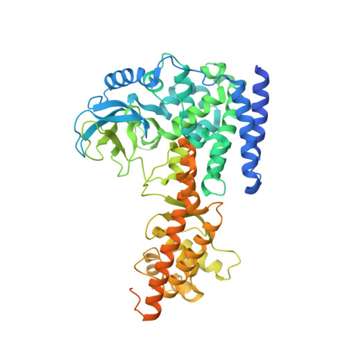
6MBJ, 6MBK, 6MBL - PubMed Abstract:
For more than 50 years, the methylation of mammalian actin at histidine 73 has been known to occur 1 . Despite the pervasiveness of His73 methylation, which we find is conserved in several model animals and plants, its function remains unclear and the enzyme that generates this modification is unknown. Here we identify SET domain protein 3 (SETD3) as the physiological actin His73 methyltransferase. Structural studies reveal that an extensive network of interactions clamps the actin peptide onto the surface of SETD3 to orient His73 correctly within the catalytic pocket and to facilitate methyl transfer. His73 methylation reduces the nucleotide-exchange rate on actin monomers and modestly accelerates the assembly of actin filaments. Mice that lack SETD3 show complete loss of actin His73 methylation in several tissues, and quantitative proteomics analysis shows that actin His73 methylation is the only detectable physiological substrate of SETD3. SETD3-deficient female mice have severely decreased litter sizes owing to primary maternal dystocia that is refractory to ecbolic induction agents. Furthermore, depletion of SETD3 impairs signal-induced contraction in primary human uterine smooth muscle cells. Together, our results identify a mammalian histidine methyltransferase and uncover a pivotal role for SETD3 and actin His73 methylation in the regulation of smooth muscle contractility. Our data also support the broader hypothesis that protein histidine methylation acts as a common regulatory mechanism.
- Department of Biology, Stanford University, Stanford, CA, USA.
Organizational Affiliation: