6M84

Crystal structure of cKir2.2 force open mutant in complex with PI(4,5)P2


Experimental Data Snapshot

  • Method: X-RAY DIFFRACTION
  • Resolution: 2.81 Å
  • R-Value Free: 0.269 
  • R-Value Work: 0.222 
  • R-Value Observed: 0.224 

wwPDB Validation   3D Report Full Report


Ligand Structure Quality Assessment 


This is version 1.3 of the entry. See complete history


Literature

Atomistic basis of opening and conduction in mammalian inward rectifier potassium (Kir2.2) channels.

Zangerl-Plessl, E.M.Lee, S.J.Maksaev, G.Bernsteiner, H.Ren, F.Yuan, P.Stary-Weinzinger, A.Nichols, C.G.

(2020) J Gen Physiol 152

  • DOI: https://doi.org/10.1085/jgp.201912422
  • Primary Citation of Related Structures:  
    6M84, 6M85

  • PubMed Abstract: 

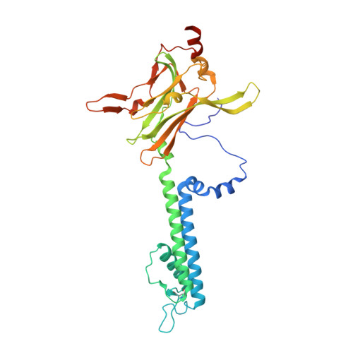
    Potassium ion conduction through open potassium channels is essential to control of membrane potentials in all cells. To elucidate the open conformation and hence the mechanism of K+ ion conduction in the classic inward rectifier Kir2.2, we introduced a negative charge (G178D) at the crossing point of the inner helix bundle, the location of ligand-dependent gating. This "forced open" mutation generated channels that were active even in the complete absence of phosphatidylinositol-4,5-bisphosphate (PIP2), an otherwise essential ligand for Kir channel opening. Crystal structures were obtained at a resolution of 3.6 Å without PIP2 bound, or 2.8 Å in complex with PIP2. The latter revealed a slight widening at the helix bundle crossing (HBC) through backbone movement. MD simulations showed that subsequent spontaneous wetting of the pore through the HBC gate region allowed K+ ion movement across the HBC and conduction through the channel. Further simulations reveal atomistic details of the opening process and highlight the role of pore-lining acidic residues in K+ conduction through Kir2 channels.


  • Organizational Affiliation

    Department of Pharmacology and Toxicology, University of Vienna, Vienna, Austria.


Macromolecules
Find similar proteins by:  (by identity cutoff)  |  3D Structure
Entity ID: 1
MoleculeChains Sequence LengthOrganismDetailsImage
ATP-sensitive inward rectifier potassium channel 12343Gallus gallusMutation(s): 2 
Gene Names: KCNJ12
Membrane Entity: Yes 
UniProt
Find proteins for F1NHE9 (Gallus gallus)
Explore F1NHE9 
Go to UniProtKB:  F1NHE9
Entity Groups  
Sequence Clusters30% Identity50% Identity70% Identity90% Identity95% Identity100% Identity
UniProt GroupF1NHE9
Sequence Annotations
Expand
  • Reference Sequence
Small Molecules
Ligands 3 Unique
IDChains Name / Formula / InChI Key2D Diagram3D Interactions
PIO
Query on PIO

Download Ideal Coordinates CCD File 
H [auth A][(2R)-2-octanoyloxy-3-[oxidanyl-[(1R,2R,3S,4R,5R,6S)-2,3,6-tris(oxidanyl)-4,5-diphosphonooxy-cyclohexyl]oxy-phosphoryl]oxy-propyl] octanoate
C25 H49 O19 P3
XLNCEHRXXWQMPK-MJUMVPIBSA-N
LMT
Query on LMT

Download Ideal Coordinates CCD File 
I [auth A]DODECYL-BETA-D-MALTOSIDE
C24 H46 O11
NLEBIOOXCVAHBD-QKMCSOCLSA-N
K
Query on K

Download Ideal Coordinates CCD File 
B [auth A]
C [auth A]
D [auth A]
E [auth A]
F [auth A]
B [auth A],
C [auth A],
D [auth A],
E [auth A],
F [auth A],
G [auth A]
POTASSIUM ION
K
NPYPAHLBTDXSSS-UHFFFAOYSA-N
Experimental Data & Validation

Experimental Data

  • Method: X-RAY DIFFRACTION
  • Resolution: 2.81 Å
  • R-Value Free: 0.269 
  • R-Value Work: 0.222 
  • R-Value Observed: 0.224 
  • Space Group: I 4
Unit Cell:
Length ( Å )Angle ( ˚ )
a = 82.787α = 90
b = 82.787β = 90
c = 182.976γ = 90
Software Package:
Software NamePurpose
REFMACrefinement
HKL-2000data reduction
SCALEPACKdata scaling
MOLREPphasing
Cootmodel building
PDB_EXTRACTdata extraction

Structure Validation

View Full Validation Report


Ligand Structure Quality Assessment 


Entry History & Funding Information

Deposition Data


Funding OrganizationLocationGrant Number
National Institutes of Health/National Human Genome Research Institute (NIH/NHGRI)United StatesHL54171
American Heart AssociationUnited States15POST22390016

Revision History  (Full details and data files)

  • Version 1.0: 2019-09-04
    Type: Initial release
  • Version 1.1: 2019-12-18
    Changes: Author supporting evidence
  • Version 1.2: 2020-09-16
    Changes: Database references, Derived calculations
  • Version 1.3: 2023-10-11
    Changes: Data collection, Database references, Refinement description