6M5J | pdb_00006m5j

U shaped head to head four-way junction in d(TTCTGCTGCTGAA/TTCTGCAGCTGAA) sequence


Experimental Data Snapshot

  • Method: X-RAY DIFFRACTION
  • Resolution: 1.65 Å
  • R-Value Free: 
    0.208 (Depositor), 0.210 (DCC) 
  • R-Value Work: 
    0.187 (Depositor), 0.190 (DCC) 
  • R-Value Observed: 
    0.188 (Depositor) 

Starting Model: experimental
View more details

wwPDB Validation   3D Report Full Report


Ligand Structure Quality Assessment 


This is version 1.1 of the entry. See complete history


Literature

Structural Basis for Targeting T:T Mismatch with Triaminotriazine-Acridine Conjugate Induces a U-Shaped Head-to-Head Four-Way Junction in CTG Repeat DNA.

Chien, C.M.Wu, P.C.Satange, R.Chang, C.C.Lai, Z.L.Hagler, L.D.Zimmerman, S.C.Hou, M.H.

(2020) J Am Chem Soc 142: 11165-11172

  • DOI: https://doi.org/10.1021/jacs.0c03591
  • Primary Citation of Related Structures:  
    6M4T, 6M5J

  • PubMed Abstract: 

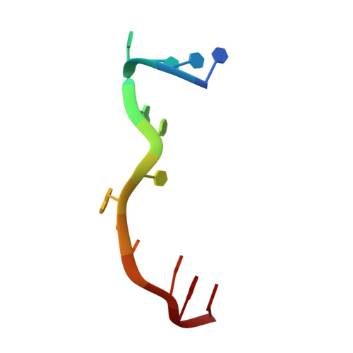
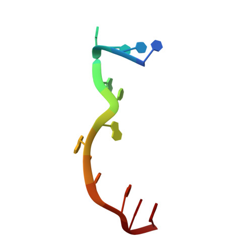
    The potent DNA-binding compound triaminotriazine-acridine conjugate (Z1) functions by targeting T:T mismatches in CTG trinucleotide repeats that are responsible for causing neurological diseases such as myotonic dystrophy type 1, but its binding mechanism remains unclear. We solved a crystal structure of Z1 in a complex with DNA containing three consecutive CTG repeats with three T:T mismatches. Crystallographic studies revealed that direct intercalation of two Z1 molecules at both ends of the CTG repeat induces thymine base flipping and DNA backbone deformation to form a four-way junction. The core of the complex unexpectedly adopts a U-shaped head-to-head topology to form a crossover of each chain at the junction site. The crossover junction is held together by two stacked G:C pairs at the central core that rotate with respect to each other in an X-shape to form two nonplanar minor-groove-aligned G·C·G·C tetrads. Two stacked G:C pairs on both sides of the center core are involved in the formation of pseudo-continuous duplex DNA. Four metal-mediated base pairs are observed between the N7 atoms of G and Co II , an interaction that strongly preserves the central junction site. Beyond revealing a new type of ligand-induced, four-way junction, these observations enhance our understanding of the specific supramolecular chemistry of Z1 that is essential for the formation of a noncanonical DNA superstructure. The structural features described here serve as a foundation for the design of new sequence-specific ligands targeting mismatches in the repeat-associated structures.


  • Organizational Affiliation

    Department of Chemistry, University of Illinois at Urbana-Champaign, Urbana, Illinois 61801, United States.


Macromolecules

Find similar nucleic acids by:  Sequence   |   3D Structure  

Entity ID: 1
MoleculeChains LengthOrganismImage
DNA (5'-D(P*TP*TP*CP*TP*GP*CP*TP*GP*CP*TP*GP*AP*A)-3')
A, C, D
13synthetic construct
Sequence Annotations
Expand
  • Reference Sequence

Find similar nucleic acids by:  Sequence   |   3D Structure  

Entity ID: 2
MoleculeChains LengthOrganismImage
DNA (5'-D(P*TP*TP*CP*TP*GP*CP*AP*GP*CP*TP*GP*AP*A)-3')13synthetic construct
Sequence Annotations
Expand
  • Reference Sequence
Small Molecules
Ligands 2 Unique
IDChains Name / Formula / InChI Key2D Diagram3D Interactions
F1R (Subject of Investigation/LOI)
Query on F1R

Download Ideal Coordinates CCD File 
G [auth A],
J [auth B],
K [auth C],
L [auth D]
N4-[4-[(6-chloranyl-2-methoxy-acridin-9-yl)amino]butyl]-1,3,5-triazine-2,4,6-triamine
C21 H23 Cl N8 O
YRQBNFDRSZQBNL-UHFFFAOYSA-N
CO (Subject of Investigation/LOI)
Query on CO

Download Ideal Coordinates CCD File 
E [auth A],
F [auth A],
H [auth B],
I [auth B]
COBALT (II) ION
Co
XLJKHNWPARRRJB-UHFFFAOYSA-N
Experimental Data & Validation

Experimental Data

  • Method: X-RAY DIFFRACTION
  • Resolution: 1.65 Å
  • R-Value Free:  0.208 (Depositor), 0.210 (DCC) 
  • R-Value Work:  0.187 (Depositor), 0.190 (DCC) 
  • R-Value Observed: 0.188 (Depositor) 
Space Group: P 1 21 1
Unit Cell:
Length ( Å )Angle ( ˚ )
a = 57.63α = 90
b = 62.086β = 121.84
c = 30.747γ = 90
Software Package:
Software NamePurpose
HKL-2000data scaling
PHENIXrefinement
PDB_EXTRACTdata extraction
HKL-2000data reduction
PHENIXphasing

Structure Validation

View Full Validation Report


Ligand Structure Quality Assessment 


Entry History 

Deposition Data

Revision History  (Full details and data files)

  • Version 1.0: 2020-07-15
    Type: Initial release
  • Version 1.1: 2023-11-29
    Changes: Advisory, Data collection, Database references, Derived calculations, Refinement description