


Crystal structure of the mouse endonuclease EndoG(H138A/C100A), space group C2
Park, K.H., Woo, E.J.To be published.
Experimental Data Snapshot
Starting Model: experimental
View more details
wwPDB Validation 3D Report Full Report

Entity ID: 1 | |||||
|---|---|---|---|---|---|
| Molecule | Chains | Sequence Length | Organism | Details | Image |
| Endonuclease G, mitochondrial | 249 | Mus musculus | Mutation(s): 2 Gene Names: Endog EC: 3.1.30 | 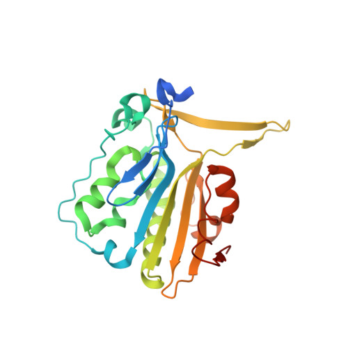 | |
UniProt & NIH Common Fund Data Resources | |||||
IMPC: MGI:1261433 | |||||
Entity Groups | |||||
| Sequence Clusters | 30% Identity50% Identity70% Identity90% Identity95% Identity100% Identity | ||||
| UniProt Group | O08600 | ||||
Sequence AnnotationsExpand | |||||
Reference Sequence | |||||
| Length ( Å ) | Angle ( ˚ ) |
|---|---|
| a = 252.407 | α = 90 |
| b = 81.34 | β = 102.17 |
| c = 54.684 | γ = 90 |
| Software Name | Purpose |
|---|---|
| PHENIX | refinement |
| HKL-2000 | data reduction |
| HKL-2000 | data scaling |
| PHENIX | phasing |


