6LUG

Crystal structure of N(omega)-hydroxy-L-arginine hydrolase


Experimental Data Snapshot

  • Method: X-RAY DIFFRACTION
  • Resolution: 1.90 Å
  • R-Value Free: 0.250 
  • R-Value Work: 0.203 
  • R-Value Observed: 0.206 

wwPDB Validation   3D Report Full Report


This is version 1.1 of the entry. See complete history

Re-refinement Note

A newer entry is available that reflects an alternative modeling of the original data: 6LUH


Literature

Crystal structure of an Nomega-hydroxy-L-arginine hydrolase found in the D-cycloserine biosynthetic pathway.

Oda, K.Shimotani, N.Kuroda, T.Matoba, Y.

(2020) Acta Crystallogr D Struct Biol 76: 506-514

  • DOI: https://doi.org/10.1107/S2059798320004908
  • Primary Citation of Related Structures:  
    6LUG, 6LUH

  • PubMed Abstract: 

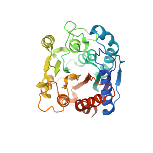
    DcsB, one of the enzymes encoded in the D-cycloserine (D-CS) biosynthetic gene cluster, displays a high sequence homology to arginase, which contains two manganese ions in the active site. However, DcsB hydrolyzes N ω -hydroxy-L-arginine, but not L-arginine, to supply hydroxyurea for the biosynthesis of D-CS. Here, the crystal structure of DcsB was determined at a resolution of 1.5 Å using anomalous scattering from the manganese ions. In the crystal structure, DscB generates an artificial dimer created by the open and closed forms. Gel-filtration analysis demonstrated that DcsB is a monomeric protein, unlike arginase, which forms a trimeric structure. The active center containing the binuclear manganese cluster differs between DcsB and arginase. In DcsB, one of the ligands of the Mn A ion is a cysteine, while the corresponding residue in arginase is a histidine. In addition, DcsB has no counterpart to the histidine residue that acts as a general acid/base during the catalytic reaction of arginase. The present study demonstrates that DcsB has a unique active site that differs from that of arginase.


  • Organizational Affiliation

    Department of Virology, Institute of Biomedical and Health Sciences, Hiroshima University, 1-2-3 Kasumi, Minami-ku, Hiroshima 734-8553, Japan.


Macromolecules
Find similar proteins by:  (by identity cutoff)  |  3D Structure
Entity ID: 1
MoleculeChains Sequence LengthOrganismDetailsImage
N(omega)-hydroxy-L-arginine amidinohydrolase
A, B
281Streptomyces lavendulaeMutation(s): 0 
Gene Names: dcsB
EC: 3.5.3.25
UniProt
Find proteins for D2Z025 (Streptomyces lavendulae)
Explore D2Z025 
Go to UniProtKB:  D2Z025
Entity Groups  
Sequence Clusters30% Identity50% Identity70% Identity90% Identity95% Identity100% Identity
UniProt GroupD2Z025
Sequence Annotations
Expand
  • Reference Sequence
Experimental Data & Validation

Experimental Data

  • Method: X-RAY DIFFRACTION
  • Resolution: 1.90 Å
  • R-Value Free: 0.250 
  • R-Value Work: 0.203 
  • R-Value Observed: 0.206 
  • Space Group: P 1
Unit Cell:
Length ( Å )Angle ( ˚ )
a = 46.44α = 83.44
b = 47.02β = 84.59
c = 59.35γ = 70.31
Software Package:
Software NamePurpose
PHENIXrefinement
XDSdata reduction
XSCALEdata scaling
SHARPphasing
PDB_EXTRACTdata extraction

Structure Validation

View Full Validation Report


Entry History 

Deposition Data

Revision History  (Full details and data files)

  • Version 1.0: 2020-06-17
    Type: Initial release
  • Version 1.1: 2024-03-27
    Changes: Data collection, Database references, Derived calculations