Thermodynamic Control of Domain Swapping by Modulating the Helical Propensity in the Hinge Region of Myoglobin.
Nagao, S., Suda, A., Kobayashi, H., Shibata, N., Higuchi, Y., Hirota, S.(2020) Chem Asian J 15: 1743-1749
- PubMed: 32329228 Search on PubMed
- DOI: https://doi.org/10.1002/asia.202000307
- Primary Citation Related Structures:
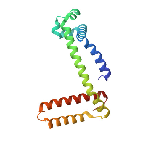
6LS8, 6LTL, 6LTM - PubMed Abstract:
Domain swapping is an exception to Anfinsen's dogma, and more than one structure can be produced from the same amino acid sequence by domain swapping. We have previously shown that myoglobin (Mb) can form a domain-swapped dimer in which the hinge region is converted to a helical structure. In this study, we showed that domain-swapped dimerization of Mb was achieved by a single Ala mutation of Gly at position 80. Multiple Ala mutations at positions 81 and 82 in addition to position 80 facilitated dimerization of Mb by stabilization of the dimeric states. Domain swapping tendencies correlated well with the helical propensity of the mutated residue in a series of Mb mutants with amino acids introduced to the hinge region. These findings demonstrate that a single mutation in the hinge loop to modify helical propensity can control oligomer formation, providing new ideas to create high-order protein oligomers using domain swapping.
- Division of Materials Science, Graduate School of Science and Technology, Nara Institute of Science and Technology, 8916-5 Takayama, Ikoma, Nara, 630-0192, Japan.
Organizational Affiliation: