


Crystal structures of Uso1 membrane tether reveal an alternative conformation in the globular head domain
Heo, Y., Yoon, H.J., Ko, H., Jang, S., Lee, H.H.(2020) Sci Rep 10: 9544
Experimental Data Snapshot
Starting Model: experimental
View more details
wwPDB Validation 3D Report Full Report

(2020) Sci Rep 10: 9544
Entity ID: 1 | |||||
|---|---|---|---|---|---|
| Molecule | Chains | Sequence Length | Organism | Details | Image |
| Intracellular protein transport protein USO1 | 731 | Saccharomyces cerevisiae S288C | Mutation(s): 0 Gene Names: USO1 | 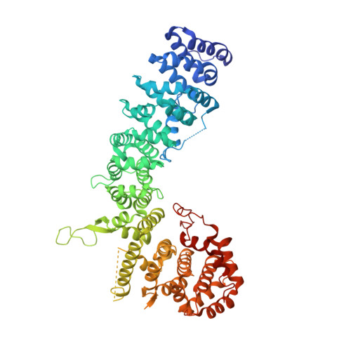 | |
UniProt | |||||
Entity Groups | |||||
| Sequence Clusters | 30% Identity50% Identity70% Identity90% Identity95% Identity100% Identity | ||||
| UniProt Group | P25386 | ||||
Sequence AnnotationsExpand | |||||
Reference Sequence | |||||
| Length ( Å ) | Angle ( ˚ ) |
|---|---|
| a = 104.376 | α = 90 |
| b = 104.376 | β = 90 |
| c = 231.842 | γ = 120 |
| Software Name | Purpose |
|---|---|
| HKL-2000 | data scaling |
| REFMAC | refinement |
| PDB_EXTRACT | data extraction |
| HKL-2000 | data reduction |
| MOLREP | phasing |


