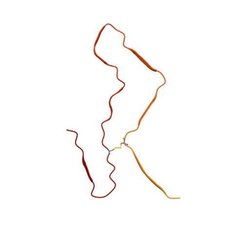
6LNI | pdb_00006lni

Cryo-EM structure of amyloid fibril formed by full-length human prion protein


Experimental Data Snapshot

  • Method: ELECTRON MICROSCOPY
  • Resolution: 2.70 Å
  • Aggregation State: HELICAL ARRAY 
  • Reconstruction Method: HELICAL 

wwPDB Validation 3D Report Full Report

Validation slider image for 6LNI

This is version 1.2 of the entry. See complete history. 

Literature

Cryo-EM structure of an amyloid fibril formed by full-length human prion protein.

Wang, L.Q., Zhao, K., Yuan, H.Y., Wang, Q., Guan, Z., Tao, J., Li, X.N., Sun, Y., Yi, C.W., Chen, J., Li, D., Zhang, D., Yin, P., Liu, C., Liang, Y.

(2020) Nat Struct Mol Biol 27: 598-602

  • DOI: https://doi.org/10.1038/s41594-020-0441-5
  • Primary Citation Related Structures: 
    6LNI

  • PubMed Abstract: 

    Prion diseases are caused by the misfolding of prion protein (PrP). Misfolded PrP forms protease-resistant aggregates in vivo (PrP Sc ) that are able to template the conversion of the native form of the protein (PrP C ), a property shared by in vitro-produced PrP fibrils. Here we produced amyloid fibrils in vitro from recombinant, full-length human PrP C (residues 23-231) and determined their structure using cryo-EM, building a model for the fibril core comprising residues 170-229. The PrP fibril consists of two protofibrils intertwined in a left-handed helix. Lys194 and Glu196 from opposing subunits form salt bridges, creating a hydrophilic cavity at the interface of the two protofibrils. By comparison with the structure of PrP C , we propose that two α-helices in the C-terminal domain of PrP C are converted into β-strands stabilized by a disulfide bond in the PrP fibril. Our data suggest that different PrP mutations may play distinct roles in modulating the conformational conversion.


  • Organizational Affiliation: 
    • Hubei Key Laboratory of Cell Homeostasis, College of Life Sciences, Wuhan University, Wuhan, China.

Macromolecule Content 

  • Total Structure Weight: 229.96 kDa 
  • Atom Count: 4,920 
  • Modeled Residue Count: 600 
  • Deposited Residue Count: 2,100 
  • Unique protein chains: 1

Macromolecules

Find similar proteins by:|  3D Structure
Entity ID: 1
MoleculeChains  Sequence LengthOrganismDetailsImage
Major prion protein
A, B, C, D, E
A, B, C, D, E, F, G, H, I, J
210Homo sapiensMutation(s): 0 
Gene Names: PRNP, ALTPRP, PRIP, PRP
UniProt & NIH Common Fund Data Resources
Find proteins for P04156 (Homo sapiens)
Explore P04156 
Go to UniProtKB:  P04156
PHAROS:  P04156
GTEx:  ENSG00000171867 
Entity Groups
Sequence Clusters30% Identity50% Identity70% Identity90% Identity95% Identity100% Identity
UniProt GroupP04156
Sequence Annotations
Expand
Reference Sequence

Experimental Data & Validation

Experimental Data

  • Method: ELECTRON MICROSCOPY
  • Resolution: 2.70 Å
  • Aggregation State: HELICAL ARRAY 
  • Reconstruction Method: HELICAL 

Structure Validation

View Full Validation Report


Entry History 

Deposition Data

Revision History  (Full details and data files)

  • Version 1.0: 2020-06-10
    Type: Initial release
  • Version 1.1: 2020-06-24
    Changes: Database references
  • Version 1.2: 2024-10-09
    Changes: Data collection, Database references, Structure summary